 Movie
Movie Controller

[English] 日本語
 Yorodumi
- PDB-5u0k: C-terminal ankyrin repeats from human liver-type glutaminase (GAB/LGA) -
+ Open data

- Basic information
| Entry | Database: PDB / ID: 5u0k | ||||||
|---|---|---|---|---|---|---|---|
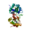
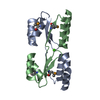
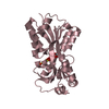
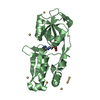
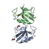
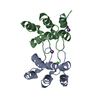
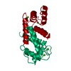
| Title | C-terminal ankyrin repeats from human liver-type glutaminase (GAB/LGA) | ||||||
 Components Components | Glutaminase liver isoform, mitochondrial | ||||||
 Keywords Keywords |  HYDROLASE / Glutaminase / ankyrin / human / GLS2 HYDROLASE / Glutaminase / ankyrin / human / GLS2 | ||||||
| Function / homology |  Function and homology information Function and homology informationglutamine catabolic process / glutamate biosynthetic process / Glutamate and glutamine metabolism / Glutamate Neurotransmitter Release Cycle / glutaminase / glutaminase activity / amino acid metabolic process / reactive oxygen species metabolic process / TP53 Regulates Metabolic Genes / regulation of apoptotic process ...glutamine catabolic process / glutamate biosynthetic process / Glutamate and glutamine metabolism / Glutamate Neurotransmitter Release Cycle / glutaminase / glutaminase activity / amino acid metabolic process / reactive oxygen species metabolic process / TP53 Regulates Metabolic Genes / regulation of apoptotic process / mitochondrial matrix / mitochondrionSimilarity search - Function | ||||||
| Biological species |  Homo sapiens (human) Homo sapiens (human) | ||||||
| Method | X-RAY DIFFRACTION / SYNCHROTRON / MOLECULAR REPLACEMENT / molecular replacement / Resolution: 2.548 Å | ||||||
 Authors Authors | Ferreira, I.M. / Pasquali, C.C. / Gonzalez, A. / Dias, S.M.G. / Ambrosio, A.L.B. | ||||||
| Funding support |  Brazil, 1items Brazil, 1items
| ||||||
 Citation Citation | Journal: J. Biol. Chem. / Year: 2017 Title: The origin and evolution of human glutaminases and their atypical C-terminal ankyrin repeats. Authors: Pasquali, C.C. / Islam, Z. / Adamoski, D. / Ferreira, I.M. / Righeto, R.D. / Bettini, J. / Portugal, R.V. / Yue, W.W. / Gonzalez, A. / Dias, S.M.G. / Ambrosio, A.L.B. | ||||||
| History |
|
- Structure visualization
| Structure viewer | Molecule: MolmilJmol/JSmol |
|---|
- Downloads & links
-Download
| PDBx/mmCIF format | 5u0k.cif.gz | 202.8 KB | Display | PDBx/mmCIF format |
|---|---|---|---|---|
| PDB format | pdb5u0k.ent.gz | 161.1 KB | Display | PDB format |
| PDBx/mmJSON format | 5u0k.json.gz | Tree view | PDBx/mmJSON format | |
| Others |  Other downloads Other downloads |
-Validation report
| Arichive directory | https://data.pdbj.org/pub/pdb/validation_reports/u0/5u0kftp://data.pdbj.org/pub/pdb/validation_reports/u0/5u0k | HTTPS FTP |
|---|
-Related structure data
| Related structure data |  5u0iSC  5u0jC  5uqeC S: Starting model for refinement C: citing same article ( |
|---|---|
| Similar structure data |


-Links
 PDBj
PDBj
- Assembly
| Deposited unit | 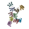
| ||||||||
|---|---|---|---|---|---|---|---|---|---|
| 1 | 
| ||||||||
| 2 | 
| ||||||||
| 3 | 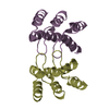
| ||||||||
| 4 | 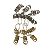
| ||||||||
| 5 | 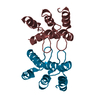
| ||||||||
| Unit cell |
|
-Components
| #1: Protein | Mass: 15840.733 Da / Num. of mol.: 10 / Fragment: UNP residues 485-602 Source method: isolated from a genetically manipulated source Source: (gene. exp.) Homo sapiens (human) / Gene: GLS2, GA / Production host:  Escherichia coli (E. coli) / Strain (production host): Rosetta 2 (DE3) / References: UniProt: Q9UI32, glutaminase Escherichia coli (E. coli) / Strain (production host): Rosetta 2 (DE3) / References: UniProt: Q9UI32, glutaminase#2: Water | ChemComp-HOH / | Water Mass: 18.015 Da / Num. of mol.: 118 / Source method: isolated from a natural source / Formula: H2O Mass: 18.015 Da / Num. of mol.: 118 / Source method: isolated from a natural source / Formula: H2O |
|---|
-Experimental details
-Experiment
| Experiment | Method: X-RAY DIFFRACTION / Number of used crystals: 1 |
|---|
- Sample preparation
| Crystal | Density Matthews: 2.23 Å3/Da / Density % sol: 44.86 % |
|---|---|
| Crystal grow | Temperature: 277 K / Method: vapor diffusion, sitting drop / pH: 8 Details: 1.1 M tri-sodium citrate, 0.1 M imidazole, 20 mM glutamine |
-Data collection
| Diffraction | Mean temperature: 100 K | |||||||||||||||||||||
|---|---|---|---|---|---|---|---|---|---|---|---|---|---|---|---|---|---|---|---|---|---|---|
| Diffraction source | Source: SYNCHROTRON / Site: SSRL  / Beamline: BL12-2 / Wavelength: 0.9795 Å / Beamline: BL12-2 / Wavelength: 0.9795 Å | |||||||||||||||||||||
| Detector | Type: DECTRIS PILATUS 6M / Detector: PIXEL / Date: May 17, 2015 / Details: Rh coated mirror | |||||||||||||||||||||
| Radiation | Monochromator: Liquid nitrogen-cooled double crystal Si(111) Protocol: SINGLE WAVELENGTH / Monochromatic (M) / Laue (L): M / Scattering type: x-ray | |||||||||||||||||||||
| Radiation wavelength | Wavelength: 0.9795 Å / Relative weight: 1 | |||||||||||||||||||||
| Reflection | Resolution: 2.55→84.2 Å / Num. obs: 45005 / % possible obs: 99.9 % / Redundancy: 8.1 % / CC1/2: 0.999 / Rmerge(I) obs: 0.094 / Net I/σ(I): 14.1 | |||||||||||||||||||||
| Reflection shell |
|
-Phasing
| Phasing | Method: molecular replacement |
|---|
- Processing
| Software |
| |||||||||||||||||||||||||||||||||||||||||||||||||||||||||||||||||||||||||||||||||||||||||||||||||||||||||||||||||||||||||||||||||||||||||||||||||||||||||||||||||||||||||||||||||||||||||||||||||||||||||||||||||||||||||
|---|---|---|---|---|---|---|---|---|---|---|---|---|---|---|---|---|---|---|---|---|---|---|---|---|---|---|---|---|---|---|---|---|---|---|---|---|---|---|---|---|---|---|---|---|---|---|---|---|---|---|---|---|---|---|---|---|---|---|---|---|---|---|---|---|---|---|---|---|---|---|---|---|---|---|---|---|---|---|---|---|---|---|---|---|---|---|---|---|---|---|---|---|---|---|---|---|---|---|---|---|---|---|---|---|---|---|---|---|---|---|---|---|---|---|---|---|---|---|---|---|---|---|---|---|---|---|---|---|---|---|---|---|---|---|---|---|---|---|---|---|---|---|---|---|---|---|---|---|---|---|---|---|---|---|---|---|---|---|---|---|---|---|---|---|---|---|---|---|---|---|---|---|---|---|---|---|---|---|---|---|---|---|---|---|---|---|---|---|---|---|---|---|---|---|---|---|---|---|---|---|---|---|---|---|---|---|---|---|---|---|---|---|---|---|---|---|---|---|
| Refinement | Method to determine structure: MOLECULAR REPLACEMENT Starting model: 5U0I Resolution: 2.548→38.033 Å / SU ML: 0.4 / Cross valid method: THROUGHOUT / σ(F): 0 / Phase error: 28.94 / Stereochemistry target values: ML
| |||||||||||||||||||||||||||||||||||||||||||||||||||||||||||||||||||||||||||||||||||||||||||||||||||||||||||||||||||||||||||||||||||||||||||||||||||||||||||||||||||||||||||||||||||||||||||||||||||||||||||||||||||||||||
| Solvent computation | Shrinkage radii: 0.9 Å / VDW probe radii: 1.11 Å / Solvent model: FLAT BULK SOLVENT MODEL | |||||||||||||||||||||||||||||||||||||||||||||||||||||||||||||||||||||||||||||||||||||||||||||||||||||||||||||||||||||||||||||||||||||||||||||||||||||||||||||||||||||||||||||||||||||||||||||||||||||||||||||||||||||||||
| Refinement step | Cycle: LAST / Resolution: 2.548→38.033 Å
| |||||||||||||||||||||||||||||||||||||||||||||||||||||||||||||||||||||||||||||||||||||||||||||||||||||||||||||||||||||||||||||||||||||||||||||||||||||||||||||||||||||||||||||||||||||||||||||||||||||||||||||||||||||||||
| Refine LS restraints |
| |||||||||||||||||||||||||||||||||||||||||||||||||||||||||||||||||||||||||||||||||||||||||||||||||||||||||||||||||||||||||||||||||||||||||||||||||||||||||||||||||||||||||||||||||||||||||||||||||||||||||||||||||||||||||
| LS refinement shell |
|
