 Movie
Movie Controller


+ Open data

- Basic information
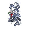
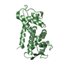
| Entry | Database: PDB / ID: 1a0o | ||||||
|---|---|---|---|---|---|---|---|
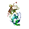
| Title | CHEY-BINDING DOMAIN OF CHEA IN COMPLEX WITH CHEY | ||||||
 Components Components |
| ||||||
 Keywords Keywords |  CHEMOTAXIS / BACTERIAL CHEMOTAXIS / SIGNAL TRANSDUCTION / TWO-COMPONENT SYSTEM / HISTIDINE KINASE / RESPONSE REGULATOR CHEMOTAXIS / BACTERIAL CHEMOTAXIS / SIGNAL TRANSDUCTION / TWO-COMPONENT SYSTEM / HISTIDINE KINASE / RESPONSE REGULATOR | ||||||
| Function / homology |  Function and homology information Function and homology informationnegative regulation of protein modification process / methyl accepting chemotaxis protein complex / positive regulation of post-translational protein modification / bacterial-type flagellum basal body, C ring / bacterial-type flagellum rotor complex / bacterial-type flagellum-dependent swimming motility / regulation of bacterial-type flagellum-dependent cell motility / aerotaxis / protein histidine kinase activity / bacterial-type flagellum ...negative regulation of protein modification process / methyl accepting chemotaxis protein complex / positive regulation of post-translational protein modification / bacterial-type flagellum basal body, C ring / bacterial-type flagellum rotor complex / bacterial-type flagellum-dependent swimming motility / regulation of bacterial-type flagellum-dependent cell motility / aerotaxis / protein histidine kinase activity / bacterial-type flagellum / regulation of chemotaxis / thermotaxis / internal peptidyl-lysine acetylation / phosphorelay response regulator activity / protein acetylation / histidine kinase / phosphorelay sensor kinase activity / acetyltransferase activity / phosphorelay signal transduction system / establishment of localization in cell / chemotaxis / phosphorylation / magnesium ion binding / signal transduction / ATP binding / plasma membrane / cytosol / cytoplasmSimilarity search - Function | ||||||
| Biological species |  Escherichia coli (E. coli) Escherichia coli (E. coli) | ||||||
| Method | X-RAY DIFFRACTION / SYNCHROTRON / MIRAS, DENSITY MODIFICATION, MOLECULAR REPLACEMENT / Resolution: 2.95 Å | ||||||
 Authors Authors | Chinardet, N. / Welch, M. / Mourey, L. / Birck, C. / Samama, J.P. | ||||||
 Citation Citation | Journal: Nat.Struct.Biol. / Year: 1998 Title: Structure of the CheY-binding domain of histidine kinase CheA in complex with CheY. Authors: Welch, M. / Chinardet, N. / Mourey, L. / Birck, C. / Samama, J.P. | ||||||
| History |
|
- Structure visualization
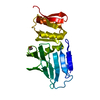
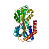
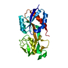
| Structure viewer | Molecule: MolmilJmol/JSmol |
|---|
- Downloads & links
-Download
| PDBx/mmCIF format | 1a0o.cif.gz | 143.6 KB | Display | PDBx/mmCIF format |
|---|---|---|---|---|
| PDB format | pdb1a0o.ent.gz | 116.8 KB | Display | PDB format |
| PDBx/mmJSON format | 1a0o.json.gz | Tree view | PDBx/mmJSON format | |
| Others |  Other downloads Other downloads |
-Validation report
| Arichive directory | https://data.pdbj.org/pub/pdb/validation_reports/a0/1a0oftp://data.pdbj.org/pub/pdb/validation_reports/a0/1a0o | HTTPS FTP |
|---|
-Related structure data
| Related structure data |  1chnS S: Starting model for refinement |
|---|---|
| Similar structure data |

-Links
 PDBj
PDBj


- Assembly
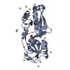
| Deposited unit | 
| ||||||||||||||||
|---|---|---|---|---|---|---|---|---|---|---|---|---|---|---|---|---|---|
| 1 | 
| ||||||||||||||||
| 2 | 
| ||||||||||||||||
| 3 | 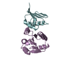
| ||||||||||||||||
| 4 | 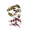
| ||||||||||||||||
| Unit cell |
| ||||||||||||||||
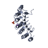
| Noncrystallographic symmetry (NCS) | NCS oper:
|
-Components
| #1: Protein | Mass: 13981.136 Da / Num. of mol.: 4 Source method: isolated from a genetically manipulated source Source: (gene. exp.) Escherichia coli (E. coli) / Cellular location: CYTOPLASM / Production host: Escherichia coli (E. coli) / References: UniProt: P06143, UniProt: P0AE67*PLUS#2: Protein | Mass: 14505.375 Da / Num. of mol.: 4 / Fragment: CHEA 124-257 Source method: isolated from a genetically manipulated source Source: (gene. exp.) Escherichia coli (E. coli) / Cellular location: CYTOPLASM / Production host: Escherichia coli (E. coli)References: UniProt: P07363, Transferases; Transferring phosphorus-containing groups; Phosphotransferases with a nitrogenous group as acceptor#3: Chemical | ChemComp-MN /   Mass: 54.938 Da / Num. of mol.: 4 / Source method: obtained synthetically / Formula: Mn Mass: 54.938 Da / Num. of mol.: 4 / Source method: obtained synthetically / Formula: Mn |
|---|
-Experimental details
-Experiment
| Experiment | Method: X-RAY DIFFRACTION / Number of used crystals: 2 |
|---|
- Sample preparation
| Crystal | Density Matthews: 2.28 Å3/Da / Density % sol: 44 % | ||||||||||||||||||||||||||||||||||||||||||||||||
|---|---|---|---|---|---|---|---|---|---|---|---|---|---|---|---|---|---|---|---|---|---|---|---|---|---|---|---|---|---|---|---|---|---|---|---|---|---|---|---|---|---|---|---|---|---|---|---|---|---|
| Crystal grow | pH: 5.5 Details: PROTEIN WAS CRYSTALLIZED FROM 20% PEG MME 5K, 0.1 M MALONIC ACID, 0.1 M MES BUFFER PH 5.5, 0.02 M DTT, 0.01 M MANGANESE CHLORIDE | ||||||||||||||||||||||||||||||||||||||||||||||||
| Crystal grow | *PLUS Temperature: 20 ℃ / pH: 7.9 / Method: vapor diffusion / Details: drop was mixed with an equal volume of reservoir | ||||||||||||||||||||||||||||||||||||||||||||||||
| Components of the solutions | *PLUS
|
-Data collection
| Diffraction | Mean temperature: 293 K |
|---|---|
| Diffraction source | Source: SYNCHROTRON / Site: LURE  / Beamline: DW32 / Wavelength: 0.97 / Beamline: DW32 / Wavelength: 0.97 |
| Detector | Type: MARRESEARCH / Detector: IMAGE PLATE / Date: Mar 1, 1996 |
| Radiation | Monochromatic (M) / Laue (L): M / Scattering type: x-ray |
| Radiation wavelength | Wavelength: 0.97 Å / Relative weight: 1 |
| Reflection | Resolution: 2.95→29 Å / Num. obs: 41612 / % possible obs: 92 % / Redundancy: 2.5 % / Rmerge(I) obs: 0.054 / Net I/σ(I): 9.5 |
| Reflection shell | Resolution: 2.95→3.03 Å / Redundancy: 2.1 % / Rmerge(I) obs: 0.099 / Mean I/σ(I) obs: 6.8 / % possible all: 86.4 |
| Reflection shell | *PLUS % possible obs: 86.4 % |
- Processing
| Software |
| ||||||||||||||||||||||||||||||||||||||||||||||||||||||||||||
|---|---|---|---|---|---|---|---|---|---|---|---|---|---|---|---|---|---|---|---|---|---|---|---|---|---|---|---|---|---|---|---|---|---|---|---|---|---|---|---|---|---|---|---|---|---|---|---|---|---|---|---|---|---|---|---|---|---|---|---|---|---|
| Refinement | Method to determine structure: MIRAS, DENSITY MODIFICATION, MOLECULAR REPLACEMENT Starting model: 1CHN Resolution: 2.95→27 Å / Rfactor Rfree error: 0.0072 / Data cutoff high absF: 100000 / Data cutoff low absF: 0.1 / Cross valid method: THROUGHOUT / σ(F): 0 Details: NCS CONSTRAINTS WERE ONLY APPLIED IN THE FIRST REFINEMENT CYCLES ONE OF THE CONSTRUCTS USED FOR THE CRYSTAL STRUCTURE DETERMINATION CONSISTS OF THE CHEY-BINDING DOMAIN OF CHEA FLANKED BY ...Details: NCS CONSTRAINTS WERE ONLY APPLIED IN THE FIRST REFINEMENT CYCLES ONE OF THE CONSTRUCTS USED FOR THE CRYSTAL STRUCTURE DETERMINATION CONSISTS OF THE CHEY-BINDING DOMAIN OF CHEA FLANKED BY DOMAIN LINKERS (CHEA124-257). IN THE FINAL MODEL, EACH MOLECULE OF THE COMPLEX COMPRISES 128 RESIDUES IN CHEY AND AN ACTIVE SITE-BOUND MN2+, AND 70 RESIDUES IN CHEA124-257 SPANNING THE REGION 159-228. NO ELECTRON DENSITY COULD BE ASSIGNED TO THE REMAINING RESIDUES IN CHEA124-257.
| ||||||||||||||||||||||||||||||||||||||||||||||||||||||||||||
| Displacement parameters | Biso mean: 33.1 Å2 | ||||||||||||||||||||||||||||||||||||||||||||||||||||||||||||
| Refine analyze |
| ||||||||||||||||||||||||||||||||||||||||||||||||||||||||||||
| Refinement step | Cycle: LAST / Resolution: 2.95→27 Å
| ||||||||||||||||||||||||||||||||||||||||||||||||||||||||||||
| Refine LS restraints |
| ||||||||||||||||||||||||||||||||||||||||||||||||||||||||||||
| Refine LS restraints NCS | NCS model details: UNRESTRAINED | ||||||||||||||||||||||||||||||||||||||||||||||||||||||||||||
| LS refinement shell | Resolution: 2.95→3.08 Å / Rfactor Rfree error: 0.032 / Total num. of bins used: 8
| ||||||||||||||||||||||||||||||||||||||||||||||||||||||||||||
| Xplor file |
| ||||||||||||||||||||||||||||||||||||||||||||||||||||||||||||
| Software | *PLUS Name: X-PLOR / Version: 3.1 / Classification: refinement | ||||||||||||||||||||||||||||||||||||||||||||||||||||||||||||
| Refine LS restraints | *PLUS
|
