 Movie
Movie Controller

[English] 日本語
 Yorodumi
- PDB-5ttk: Crystal Structure of Selenomethionine-incorporated Nicotine Oxido... -
+ Open data

- Basic information
| Entry | Database: PDB / ID: 5ttk | |||||||||
|---|---|---|---|---|---|---|---|---|---|---|
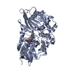
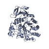
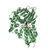
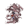
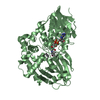
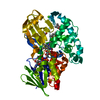
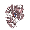
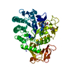
| Title | Crystal Structure of Selenomethionine-incorporated Nicotine Oxidoreductase from Pseudomonas putida | |||||||||
 Components Components | Amine oxidase | |||||||||
 Keywords Keywords | OXIDOREDUCTASE / nicotine degradation / flavoenzyme / monoamine oxidase family / PHBH | |||||||||
| Function / homology |  Function and homology informationnicotine dehydrogenase / nicotine catabolic process / alkaloid metabolic process / periplasmic space / oxidoreductase activity / nucleotide binding Function and homology informationnicotine dehydrogenase / nicotine catabolic process / alkaloid metabolic process / periplasmic space / oxidoreductase activity / nucleotide bindingSimilarity search - Function | |||||||||
| Biological species |  Pseudomonas putida (bacteria) Pseudomonas putida (bacteria) | |||||||||
| Method | X-RAY DIFFRACTION / SYNCHROTRON / SAD / Resolution: 2.51 Å | |||||||||
 Authors Authors | Tararina, M.A. / Janda, K.D. / Allen, K.N. | |||||||||
| Funding support |  United States, 2items United States, 2items
| |||||||||
 Citation Citation | Journal: Biochemistry / Year: 2016 Title: Structural Analysis Provides Mechanistic Insight into Nicotine Oxidoreductase from Pseudomonas putida. Authors: Tararina, M.A. / Janda, K.D. / Allen, K.N. | |||||||||
| History |
|
- Structure visualization
| Structure viewer | Molecule: MolmilJmol/JSmol |
|---|
- Downloads & links
-Download
| PDBx/mmCIF format | 5ttk.cif.gz | 344.1 KB | Display | PDBx/mmCIF format |
|---|---|---|---|---|
| PDB format | pdb5ttk.ent.gz | 292.7 KB | Display | PDB format |
| PDBx/mmJSON format | 5ttk.json.gz | Tree view | PDBx/mmJSON format | |
| Others |  Other downloads Other downloads |
-Validation report
| Arichive directory | https://data.pdbj.org/pub/pdb/validation_reports/tt/5ttkftp://data.pdbj.org/pub/pdb/validation_reports/tt/5ttk | HTTPS FTP |
|---|
-Related structure data


-Links
 PDBj
PDBj


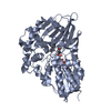
- Assembly
| Deposited unit | 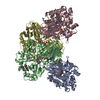
| ||||||||
|---|---|---|---|---|---|---|---|---|---|
| 1 | 
| ||||||||
| 2 | 
| ||||||||
| 3 | 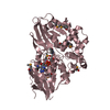
| ||||||||
| 4 | 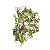
| ||||||||
| Unit cell |
|
-Components
| #1: Protein | Mass: 54134.340 Da / Num. of mol.: 4 Source method: isolated from a genetically manipulated source Source: (gene. exp.) Pseudomonas putida (strain S16) (bacteria)Strain: S16 / Gene: PPS_4081 Production host: Escherichia coli 'BL21-Gold(DE3)pLysS AG' (bacteria)References: UniProt: F8G0P2 #2: Chemical | ChemComp-FAD / Flavin adenine dinucleotide  Mass: 785.550 Da / Num. of mol.: 4 / Source method: obtained synthetically / Formula: C27H33N9O15P2 / Comment: FAD*YM Mass: 785.550 Da / Num. of mol.: 4 / Source method: obtained synthetically / Formula: C27H33N9O15P2 / Comment: FAD*YM#3: Water | ChemComp-HOH / | Water Mass: 18.015 Da / Num. of mol.: 235 / Source method: isolated from a natural source / Formula: H2O Mass: 18.015 Da / Num. of mol.: 235 / Source method: isolated from a natural source / Formula: H2O |
|---|
-Experimental details
-Experiment
| Experiment | Method: X-RAY DIFFRACTION / Number of used crystals: 1 |
|---|
- Sample preparation
| Crystal | Density Matthews: 2.41 Å3/Da / Density % sol: 48.95 % Description: three-dimensional yellow crystals of rod-like morphology with final dimensions of 0.1 mm by 0.2 mm by 0.5 mm |
|---|---|
| Crystal grow | Temperature: 290 K / Method: vapor diffusion, hanging drop / pH: 8.3 Details: 0.1 M sodium citrate tribasic dihydrate pH 8.3, 15% w/v PEG 3,350 |
-Data collection
| Diffraction | Mean temperature: 100 K |
|---|---|
| Diffraction source | Source: SYNCHROTRON / Site: SSRL / Beamline: BL12-2 / Wavelength: 0.97934 Å |
| Detector | Type: DECTRIS PILATUS 6M / Detector: PIXEL / Date: May 15, 2016 |
| Radiation | Protocol: SINGLE WAVELENGTH / Monochromatic (M) / Laue (L): M / Scattering type: x-ray |
| Radiation wavelength | Wavelength: 0.97934 Å / Relative weight: 1 |
| Reflection | Resolution: 2.51→48.551 Å / Num. obs: 69981 / % possible obs: 96 % / Redundancy: 5.3 % / CC1/2: 0.98 / Rmerge(I) obs: 0.193 / Net I/σ(I): 13.4 |
| Reflection shell | Resolution: 2.51→2.6 Å / Redundancy: 3.6 % / Rmerge(I) obs: 0.597 / Mean I/σ(I) obs: 2.6 / CC1/2: 0.88 / % possible all: 88 |
- Processing
| Software |
| |||||||||||||||||||||||||||||||||||||||||||||||||||||||||||||||||||||||||||||||||||||||||||||||||||||||||
|---|---|---|---|---|---|---|---|---|---|---|---|---|---|---|---|---|---|---|---|---|---|---|---|---|---|---|---|---|---|---|---|---|---|---|---|---|---|---|---|---|---|---|---|---|---|---|---|---|---|---|---|---|---|---|---|---|---|---|---|---|---|---|---|---|---|---|---|---|---|---|---|---|---|---|---|---|---|---|---|---|---|---|---|---|---|---|---|---|---|---|---|---|---|---|---|---|---|---|---|---|---|---|---|---|---|---|
| Refinement | Method to determine structure: SAD / Resolution: 2.51→48.551 Å / SU ML: 0.3 / Cross valid method: FREE R-VALUE / σ(F): 1.55 / Phase error: 23.79 / Stereochemistry target values: MLHL
| |||||||||||||||||||||||||||||||||||||||||||||||||||||||||||||||||||||||||||||||||||||||||||||||||||||||||
| Solvent computation | Shrinkage radii: 0.9 Å / VDW probe radii: 1.11 Å / Solvent model: FLAT BULK SOLVENT MODEL | |||||||||||||||||||||||||||||||||||||||||||||||||||||||||||||||||||||||||||||||||||||||||||||||||||||||||
| Refinement step | Cycle: LAST / Resolution: 2.51→48.551 Å
| |||||||||||||||||||||||||||||||||||||||||||||||||||||||||||||||||||||||||||||||||||||||||||||||||||||||||
| Refine LS restraints |
| |||||||||||||||||||||||||||||||||||||||||||||||||||||||||||||||||||||||||||||||||||||||||||||||||||||||||
| LS refinement shell |
|
