 Movie
Movie Controller

[English] 日本語
 Yorodumi
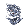
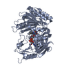
- PDB-2bvh: Crystal structure of 6-hydoxy-D-nicotine oxidase from Arthrobacte... -
+ Open data

- Basic information
| Entry | Database: PDB / ID: 2bvh | ||||||
|---|---|---|---|---|---|---|---|
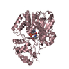
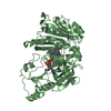
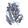
| Title | Crystal structure of 6-hydoxy-D-nicotine oxidase from Arthrobacter nicotinovorans. Crystal Form 2 (P21) | ||||||
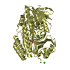
 Components Components | 6-HYDROXY-D-NICOTINE OXIDASE | ||||||
 Keywords Keywords |  OXIDASE / AUTOFLAVINYLATION / ENANTIOMERIC SUBSTRATES / FLAVOENZYMES / NICOTINE DEGRADATION OXIDASE / AUTOFLAVINYLATION / ENANTIOMERIC SUBSTRATES / FLAVOENZYMES / NICOTINE DEGRADATION | ||||||
| Function / homology |  Function and homology information(R)-6-hydroxynicotine oxidase / (R)-6-hydroxynicotine oxidase activity / nicotine catabolic process / alkaloid metabolic process / FAD binding / cytoplasm Function and homology information(R)-6-hydroxynicotine oxidase / (R)-6-hydroxynicotine oxidase activity / nicotine catabolic process / alkaloid metabolic process / FAD binding / cytoplasmSimilarity search - Function | ||||||
| Biological species |  ARTHROBACTER NICOTINOVORANS (bacteria) ARTHROBACTER NICOTINOVORANS (bacteria) | ||||||
| Method | X-RAY DIFFRACTION / SYNCHROTRON / MOLECULAR REPLACEMENT / Resolution: 2.9 Å | ||||||
 Authors Authors | Koetter, J.W.A. / Schulz, G.E. | ||||||
 Citation Citation | Journal: J.Mol.Biol. / Year: 2005 Title: Crystal Structure of 6-Hydroxy-D-Nicotine Oxidase from Arthrobacter Nicotinovorans. Authors: Koetter, J.W.A. / Schulz, G.E. | ||||||
| History |
| ||||||
| Remark 700 | SHEET THE SHEET STRUCTURE OF THIS MOLECULE IS BIFURCATED. IN ORDER TO REPRESENT THIS FEATURE IN ... SHEET THE SHEET STRUCTURE OF THIS MOLECULE IS BIFURCATED. IN ORDER TO REPRESENT THIS FEATURE IN THE SHEET RECORDS BELOW, TWO SHEETS ARE DEFINED. |
- Structure visualization
| Structure viewer | Molecule: MolmilJmol/JSmol |
|---|
- Downloads & links
-Download
| PDBx/mmCIF format | 2bvh.cif.gz | 337 KB | Display | PDBx/mmCIF format |
|---|---|---|---|---|
| PDB format | pdb2bvh.ent.gz | 286.4 KB | Display | PDB format |
| PDBx/mmJSON format | 2bvh.json.gz | Tree view | PDBx/mmJSON format | |
| Others |  Other downloads Other downloads |
-Validation report
| Arichive directory | https://data.pdbj.org/pub/pdb/validation_reports/bv/2bvhftp://data.pdbj.org/pub/pdb/validation_reports/bv/2bvh | HTTPS FTP |
|---|
-Related structure data


-Links
 PDBj
PDBj
- Assembly
| Deposited unit | 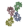
| ||||||||||||||||||||
|---|---|---|---|---|---|---|---|---|---|---|---|---|---|---|---|---|---|---|---|---|---|
| 1 | 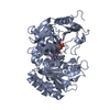
| ||||||||||||||||||||
| 2 | 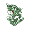
| ||||||||||||||||||||
| 3 | 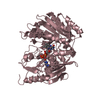
| ||||||||||||||||||||
| 4 | 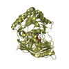
| ||||||||||||||||||||
| Unit cell |
| ||||||||||||||||||||
| Noncrystallographic symmetry (NCS) | NCS oper:
|
-Components
| #1: Protein | Mass: 48918.074 Da / Num. of mol.: 4 / Mutation: YES Source method: isolated from a genetically manipulated source Details: FAD COVALENTLY BOUND TO H 72 / Source: (gene. exp.) ARTHROBACTER NICOTINOVORANS (bacteria) / Plasmid: PKK223-3 / Production host: ESCHERICHIA COLI (E. coli) / Strain (production host): BL21(DE3) / References: UniProt: Q8GAG1, (R)-6-hydroxynicotine oxidase#2: Chemical | ChemComp-FAD / Flavin adenine dinucleotide  Mass: 785.550 Da / Num. of mol.: 4 / Source method: obtained synthetically / Formula: C27H33N9O15P2 / Comment: FAD*YM Mass: 785.550 Da / Num. of mol.: 4 / Source method: obtained synthetically / Formula: C27H33N9O15P2 / Comment: FAD*YMCompound details | ENGINEERED RESIDUE IN CHAIN A, CYS 433 TO SER ENGINEERED RESIDUE IN CHAIN B, CYS 433 TO SER ...ENGINEERED | Sequence details | MUTATION C433S | |
|---|
-Experimental details
-Experiment
| Experiment | Method: X-RAY DIFFRACTION |
|---|
- Sample preparation
| Crystal | Density Matthews: 2.5 Å3/Da / Density % sol: 50.36 % |
|---|
-Data collection
| Diffraction | Mean temperature: 100 K |
|---|---|
| Diffraction source | Source: SYNCHROTRON / Site: SLS  / Beamline: X06SA / Wavelength: 0.9787 / Beamline: X06SA / Wavelength: 0.9787 |
| Radiation | Protocol: SINGLE WAVELENGTH / Monochromatic (M) / Laue (L): M / Scattering type: x-ray |
| Radiation wavelength | Wavelength: 0.9787 Å / Relative weight: 1 |
| Reflection | Resolution: 2.9→44.3 Å / Num. obs: 42084 / % possible obs: 98.9 % / Observed criterion σ(I): 4 / Redundancy: 3.1 % / Biso Wilson estimate: 38.7 Å2 / Rmerge(I) obs: 0.1 / Net I/σ(I): 12.7 |
| Reflection shell | Resolution: 2.89→3.06 Å / Redundancy: 3 % / Rmerge(I) obs: 0.32 / Mean I/σ(I) obs: 6.2 / % possible all: 95.4 |
- Processing
| Software |
| ||||||||||||||||||||||||||||||||||||||||||||||||||||||||||||
|---|---|---|---|---|---|---|---|---|---|---|---|---|---|---|---|---|---|---|---|---|---|---|---|---|---|---|---|---|---|---|---|---|---|---|---|---|---|---|---|---|---|---|---|---|---|---|---|---|---|---|---|---|---|---|---|---|---|---|---|---|---|
| Refinement | Method to determine structure: MOLECULAR REPLACEMENT / Resolution: 2.9→44.3 Å / Rfactor Rfree error: 0.006 / Data cutoff high absF: 2955351.72 / Cross valid method: THROUGHOUT / σ(F): 0 / Stereochemistry target values: MAXIMUM LIKELIHOOD
| ||||||||||||||||||||||||||||||||||||||||||||||||||||||||||||
| Solvent computation | Solvent model: CNS BULK SOLVENT MODEL USED / Bsol: 14.437 Å2 / ksol: 0.290134 e/Å3 | ||||||||||||||||||||||||||||||||||||||||||||||||||||||||||||
| Displacement parameters | Biso mean: 47.61 Å2
| ||||||||||||||||||||||||||||||||||||||||||||||||||||||||||||
| Refine analyze |
| ||||||||||||||||||||||||||||||||||||||||||||||||||||||||||||
| Refinement step | Cycle: LAST / Resolution: 2.9→44.3 Å
| ||||||||||||||||||||||||||||||||||||||||||||||||||||||||||||
| Refine LS restraints |
|
