 Movie
Movie Controller

[English] 日本語
 Yorodumi
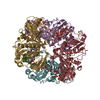
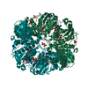
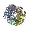
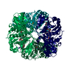
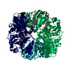
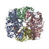
- PDB-5tso: CRYSTAL STRUCTURE OF GLYCERALDEHYDE-3-PHOSPHATE DEHYDROGENASE FRO... -
+ Open data

- Basic information
| Entry | Database: PDB / ID: 5tso | ||||||
|---|---|---|---|---|---|---|---|
| Title | CRYSTAL STRUCTURE OF GLYCERALDEHYDE-3-PHOSPHATE DEHYDROGENASE FROM PIG MUSCLE COMPLEXED WITH ORTHOPHENANTHROLINE AT 1.90 ANGSTROM RESOLUTION | ||||||
 Components Components | Glyceraldehyde-3-phosphate dehydrogenase Glyceraldehyde 3-phosphate dehydrogenase Glyceraldehyde 3-phosphate dehydrogenase | ||||||
 Keywords Keywords | OXIDOREDUCTASE / ROSSMANN FOLD / NAD / GAPDH / GLYCOLYSIS / 1 / 10-ORTHOPHENANTHROLINE | ||||||
| Function / homology |  Function and homology informationGlycolysis / peptidyl-cysteine S-trans-nitrosylation / Transferases; Transferring nitrogenous groups; Transferring other nitrogenous groups / peptidyl-cysteine S-nitrosylase activity / Gluconeogenesis / killing by host of symbiont cells / glyceraldehyde-3-phosphate dehydrogenase (phosphorylating) / negative regulation of endopeptidase activity / glyceraldehyde-3-phosphate dehydrogenase (NAD+) (phosphorylating) activity / aspartic-type endopeptidase inhibitor activity ...Glycolysis / peptidyl-cysteine S-trans-nitrosylation / Transferases; Transferring nitrogenous groups; Transferring other nitrogenous groups / peptidyl-cysteine S-nitrosylase activity / Gluconeogenesis / killing by host of symbiont cells / glyceraldehyde-3-phosphate dehydrogenase (phosphorylating) / negative regulation of endopeptidase activity / glyceraldehyde-3-phosphate dehydrogenase (NAD+) (phosphorylating) activity / aspartic-type endopeptidase inhibitor activity / GAIT complex / positive regulation of type I interferon production / defense response to fungus / lipid droplet / glycolytic process / microtubule cytoskeleton organization / cellular response to type II interferon / glucose metabolic process / NAD binding / microtubule cytoskeleton / disordered domain specific binding / antimicrobial humoral immune response mediated by antimicrobial peptide / NADP binding / microtubule binding / nuclear membrane / neuron apoptotic process / positive regulation of canonical NF-kappaB signal transduction / killing of cells of another organism / negative regulation of translation / protein stabilization / ribonucleoprotein complex / identical protein binding / nucleus / plasma membrane / cytosol / cytoplasm Function and homology informationGlycolysis / peptidyl-cysteine S-trans-nitrosylation / Transferases; Transferring nitrogenous groups; Transferring other nitrogenous groups / peptidyl-cysteine S-nitrosylase activity / Gluconeogenesis / killing by host of symbiont cells / glyceraldehyde-3-phosphate dehydrogenase (phosphorylating) / negative regulation of endopeptidase activity / glyceraldehyde-3-phosphate dehydrogenase (NAD+) (phosphorylating) activity / aspartic-type endopeptidase inhibitor activity ...Glycolysis / peptidyl-cysteine S-trans-nitrosylation / Transferases; Transferring nitrogenous groups; Transferring other nitrogenous groups / peptidyl-cysteine S-nitrosylase activity / Gluconeogenesis / killing by host of symbiont cells / glyceraldehyde-3-phosphate dehydrogenase (phosphorylating) / negative regulation of endopeptidase activity / glyceraldehyde-3-phosphate dehydrogenase (NAD+) (phosphorylating) activity / aspartic-type endopeptidase inhibitor activity / GAIT complex / positive regulation of type I interferon production / defense response to fungus / lipid droplet / glycolytic process / microtubule cytoskeleton organization / cellular response to type II interferon / glucose metabolic process / NAD binding / microtubule cytoskeleton / disordered domain specific binding / antimicrobial humoral immune response mediated by antimicrobial peptide / NADP binding / microtubule binding / nuclear membrane / neuron apoptotic process / positive regulation of canonical NF-kappaB signal transduction / killing of cells of another organism / negative regulation of translation / protein stabilization / ribonucleoprotein complex / identical protein binding / nucleus / plasma membrane / cytosol / cytoplasmSimilarity search - Function | ||||||
| Biological species |  Sus scrofa (pig) Sus scrofa (pig) | ||||||
| Method | X-RAY DIFFRACTION / SYNCHROTRON / MOLECULAR REPLACEMENT / Resolution: 1.9 Å | ||||||
 Authors Authors | Dimova, M. / Devedjiev, Y.D. | ||||||
 Citation Citation | Journal: To Be Published Title: Novel Enhancer Binding Site Found In Bacteria And Eukaryota But Not In Archea. Authors: Dimova, M. / Devedjiev, Y.D. #1: Journal: J Mol Biol. / Year: 1988Title: Coenzyme-induced conformational changes in glyceraldehyde-3-phosphate dehydrogenase from Bacillus stearothermophilus Authors: Skarzynski, T. / Wonacott, A.J. | ||||||
| History |
|
- Structure visualization
| Structure viewer | Molecule: MolmilJmol/JSmol |
|---|
- Downloads & links
-Download
| PDBx/mmCIF format | 5tso.cif.gz | 435.1 KB | Display | PDBx/mmCIF format |
|---|---|---|---|---|
| PDB format | pdb5tso.ent.gz | 358.1 KB | Display | PDB format |
| PDBx/mmJSON format | 5tso.json.gz | Tree view | PDBx/mmJSON format | |
| Others |  Other downloads Other downloads |
-Validation report
| Arichive directory | https://data.pdbj.org/pub/pdb/validation_reports/ts/5tsoftp://data.pdbj.org/pub/pdb/validation_reports/ts/5tso | HTTPS FTP |
|---|
-Related structure data
| Related structure data |  1gd1S S: Starting model for refinement |
|---|---|
| Similar structure data |


-Links
 PDBj
PDBj
- Assembly
| Deposited unit | 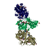
| ||||||||
|---|---|---|---|---|---|---|---|---|---|
| 1 | 
| ||||||||
| 2 | 
| ||||||||
| Unit cell |
| ||||||||
| Components on special symmetry positions |
|
-Components
-Protein , 1 types, 3 molecules PRS
| #1: Protein | Glyceraldehyde 3-phosphate dehydrogenase / GAPDH / Peptidyl-cysteine S-nitrosylase GAPDH Mass: 35883.016 Da / Num. of mol.: 3 / Source method: isolated from a natural source / Source: (natural) Sus scrofa (pig)References: UniProt: P00355, glyceraldehyde-3-phosphate dehydrogenase (phosphorylating), Transferases; Transferring nitrogenous groups; Transferring other nitrogenous groups |
|---|
-Non-polymers , 5 types, 908 molecules 


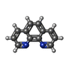

| #2: Chemical | ChemComp-SO4 / Sulfate Mass: 96.063 Da / Num. of mol.: 11 / Source method: obtained synthetically / Formula: SO4 Mass: 96.063 Da / Num. of mol.: 11 / Source method: obtained synthetically / Formula: SO4#3: Chemical | Nicotinamide adenine dinucleotide Mass: 663.425 Da / Num. of mol.: 3 / Source method: obtained synthetically / Formula: C21H27N7O14P2 / Comment: NAD*YM Mass: 663.425 Da / Num. of mol.: 3 / Source method: obtained synthetically / Formula: C21H27N7O14P2 / Comment: NAD*YM#4: Chemical | ChemComp-GOL / Glycerol Mass: 92.094 Da / Num. of mol.: 7 / Source method: obtained synthetically / Formula: C3H8O3 Mass: 92.094 Da / Num. of mol.: 7 / Source method: obtained synthetically / Formula: C3H8O3#5: Chemical | ChemComp-PHN / 1,10-Phenanthroline Mass: 180.205 Da / Num. of mol.: 4 / Source method: obtained synthetically / Formula: C12H8N2 Mass: 180.205 Da / Num. of mol.: 4 / Source method: obtained synthetically / Formula: C12H8N2#6: Water | ChemComp-HOH / | WaterMass: 18.015 Da / Num. of mol.: 883 / Source method: isolated from a natural source / Formula: H2O |
|---|
-Experimental details
-Experiment
| Experiment | Method: X-RAY DIFFRACTION / Number of used crystals: 1 |
|---|
- Sample preparation
| Crystal | Density Matthews: 3.03 Å3/Da / Density % sol: 59.38 % / Description: orthorhombic plates |
|---|---|
| Crystal grow | Temperature: 273 K / Method: vapor diffusion, hanging drop / pH: 8 Details: 0.005M 1,10-ORTHOPHENANTHROLINE, 0.05M HEPES, PH 8.0, 54.7%(W/V)AMMONIUM SULFATE,0.005 M EDTA, HANGING DROP, TEMPERATURE 277 K PH range: 8 |
-Data collection
| Diffraction | Mean temperature: 120 K |
|---|---|
| Diffraction source | Source: SYNCHROTRON / Site: APS  / Beamline: 17-ID / Wavelength: 1 Å / Beamline: 17-ID / Wavelength: 1 Å |
| Detector | Type: DECTRIS PILATUS3 2M / Detector: PIXEL / Date: Apr 15, 2005 |
| Radiation | Monochromator: DOUBLE CRYSTAL MONOCHROMATOR WITH SAGITTAL FOCUSSING Protocol: SINGLE WAVELENGTH / Monochromatic (M) / Laue (L): M / Scattering type: x-ray |
| Radiation wavelength | Wavelength: 1 Å / Relative weight: 1 |
| Reflection | Resolution: 1.9→43.54 Å / Num. obs: 87846 / % possible obs: 93.6 % / Redundancy: 4.3 % / Rmerge(I) obs: 0.066 / Net I/σ(I): 12 |
| Reflection shell | Resolution: 1.9→1.95 Å / Redundancy: 2.7 % / Rmerge(I) obs: 0.159 / % possible all: 93.6 |
- Processing
| Software |
| ||||||||||||||||||||||||||||||||||||||||||||||||||||||||||||||||||||||||||||||||||||||||||||||||||||||||||||||||||||||||||||||||||||||||||||||||||||||||||||||||||||||||||||||||||||||
|---|---|---|---|---|---|---|---|---|---|---|---|---|---|---|---|---|---|---|---|---|---|---|---|---|---|---|---|---|---|---|---|---|---|---|---|---|---|---|---|---|---|---|---|---|---|---|---|---|---|---|---|---|---|---|---|---|---|---|---|---|---|---|---|---|---|---|---|---|---|---|---|---|---|---|---|---|---|---|---|---|---|---|---|---|---|---|---|---|---|---|---|---|---|---|---|---|---|---|---|---|---|---|---|---|---|---|---|---|---|---|---|---|---|---|---|---|---|---|---|---|---|---|---|---|---|---|---|---|---|---|---|---|---|---|---|---|---|---|---|---|---|---|---|---|---|---|---|---|---|---|---|---|---|---|---|---|---|---|---|---|---|---|---|---|---|---|---|---|---|---|---|---|---|---|---|---|---|---|---|---|---|---|---|
| Refinement | Method to determine structure: MOLECULAR REPLACEMENT Starting model: 1GD1 Resolution: 1.9→43.54 Å / Cor.coef. Fo:Fc: 0.979 / Cor.coef. Fo:Fc free: 0.953 / SU B: 7.095 / SU ML: 0.09 / Cross valid method: THROUGHOUT / ESU R: 0.252 / ESU R Free: 0.123 / Stereochemistry target values: MAXIMUM LIKELIHOOD / Details: HYDROGENS HAVE BEEN ADDED IN THE RIDING POSITIONS
| ||||||||||||||||||||||||||||||||||||||||||||||||||||||||||||||||||||||||||||||||||||||||||||||||||||||||||||||||||||||||||||||||||||||||||||||||||||||||||||||||||||||||||||||||||||||
| Solvent computation | Ion probe radii: 0.8 Å / Shrinkage radii: 0.8 Å / VDW probe radii: 1.2 Å / Solvent model: MASK | ||||||||||||||||||||||||||||||||||||||||||||||||||||||||||||||||||||||||||||||||||||||||||||||||||||||||||||||||||||||||||||||||||||||||||||||||||||||||||||||||||||||||||||||||||||||
| Displacement parameters | Biso mean: 35.4 Å2
| ||||||||||||||||||||||||||||||||||||||||||||||||||||||||||||||||||||||||||||||||||||||||||||||||||||||||||||||||||||||||||||||||||||||||||||||||||||||||||||||||||||||||||||||||||||||
| Refinement step | Cycle: LAST / Resolution: 1.9→43.54 Å
| ||||||||||||||||||||||||||||||||||||||||||||||||||||||||||||||||||||||||||||||||||||||||||||||||||||||||||||||||||||||||||||||||||||||||||||||||||||||||||||||||||||||||||||||||||||||
| Refine LS restraints |
|