 Movie
Movie Controller


+ Open data

- Basic information
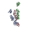
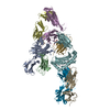
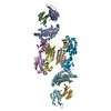
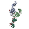
| Entry | Database: PDB / ID: 5tpn | ||||||
|---|---|---|---|---|---|---|---|
| Title | Crystal structure of RSV F in complex with human antibody hRSV90 | ||||||
 Components Components |
| ||||||
 Keywords Keywords |  IMMUNE SYSTEM / fusion protein / respiratory syncytial virus / antibody IMMUNE SYSTEM / fusion protein / respiratory syncytial virus / antibody | ||||||
| Function / homology |  Function and homology information Function and homology informationpositive regulation of syncytium formation by virus / host cell Golgi membrane / entry receptor-mediated virion attachment to host cell / symbiont entry into host cell / fusion of virus membrane with host plasma membrane / viral envelope / host cell plasma membrane / virion membrane / membrane / identical protein bindingSimilarity search - Function | ||||||
| Biological species |  Human respiratory syncytial virus AEnterobacteria phage Ox2 (virus) Human respiratory syncytial virus AEnterobacteria phage Ox2 (virus) Homo sapiens (human) Homo sapiens (human) | ||||||
| Method | X-RAY DIFFRACTION / SYNCHROTRON / MOLECULAR REPLACEMENT / Resolution: 3.141 Å | ||||||
 Authors Authors | Mousa, J.J. / Crowe, J.E. | ||||||
 Citation Citation | Journal: To Be Published Title: Crystal structure of RSV F in complex with human antibody hRSV90 Authors: Mousa, J.J. / Crowe, J.E. | ||||||
| History |
|
- Structure visualization
| Structure viewer | Molecule: MolmilJmol/JSmol |
|---|
- Downloads & links
-Download
| PDBx/mmCIF format | 5tpn.cif.gz | 187.9 KB | Display | PDBx/mmCIF format |
|---|---|---|---|---|
| PDB format | pdb5tpn.ent.gz | 147 KB | Display | PDB format |
| PDBx/mmJSON format | 5tpn.json.gz | Tree view | PDBx/mmJSON format | |
| Others |  Other downloads Other downloads |
-Validation report
| Arichive directory | https://data.pdbj.org/pub/pdb/validation_reports/tp/5tpnftp://data.pdbj.org/pub/pdb/validation_reports/tp/5tpn | HTTPS FTP |
|---|
-Related structure data


-Links
 PDBj
PDBj


- Assembly
| Deposited unit | 
| ||||||||
|---|---|---|---|---|---|---|---|---|---|
| 1 |
| ||||||||
| Unit cell |
|
-Components
| #1: Protein | Mass: 56137.848 Da / Num. of mol.: 1 / Mutation: N67I, S215P, I379V, M447V Source method: isolated from a genetically manipulated source Source: (gene. exp.) Human respiratory syncytial virus A, (gene. exp.) Enterobacteria phage Ox2 (virus)Strain: A2 / Gene: wac / Production host: Homo sapiens (human) / References: UniProt: P03420, UniProt: Q38650 |
|---|---|
| #2: Antibody | Mass: 24236.041 Da / Num. of mol.: 1 / Source method: isolated from a natural source / Source: (natural) Homo sapiens (human) |
| #3: Antibody | Mass: 23302.822 Da / Num. of mol.: 1 / Source method: isolated from a natural source / Source: (natural) Homo sapiens (human) |
-Experimental details
-Experiment
| Experiment | Method: X-RAY DIFFRACTION / Number of used crystals: 1 |
|---|
- Sample preparation
| Crystal | Density Matthews: 5.53 Å3/Da / Density % sol: 77.75 % |
|---|---|
| Crystal grow | Temperature: 293 K / Method: vapor diffusion, sitting drop Details: 30% PEG, 400/200 mM MgCl2, 6H2O/100 mM HEPES pH 7.5 |
-Data collection
| Diffraction | Mean temperature: 80 K |
|---|---|
| Diffraction source | Source: SYNCHROTRON / Site: APS  / Beamline: 21-ID-G / Wavelength: 1.0331 Å / Beamline: 21-ID-G / Wavelength: 1.0331 Å |
| Detector | Type: MARMOSAIC 300 mm CCD / Detector: CCD / Date: Apr 26, 2016 |
| Radiation | Protocol: SINGLE WAVELENGTH / Monochromatic (M) / Laue (L): M / Scattering type: x-ray |
| Radiation wavelength | Wavelength: 1.0331 Å / Relative weight: 1 |
| Reflection | Resolution: 3.1→49.289 Å / Num. obs: 30527 / % possible obs: 75.2 % / Redundancy: 4.4 % / Rmerge(I) obs: 0.28 / Net I/σ(I): 6.7 |
- Processing
| Software |
| ||||||||||||||||||||||||||||||||||||||||||||||||||||||||||||||||||||||||||||||||||||
|---|---|---|---|---|---|---|---|---|---|---|---|---|---|---|---|---|---|---|---|---|---|---|---|---|---|---|---|---|---|---|---|---|---|---|---|---|---|---|---|---|---|---|---|---|---|---|---|---|---|---|---|---|---|---|---|---|---|---|---|---|---|---|---|---|---|---|---|---|---|---|---|---|---|---|---|---|---|---|---|---|---|---|---|---|---|
| Refinement | Method to determine structure: MOLECULAR REPLACEMENT Starting model: 5C6B, 4Q9Q Resolution: 3.141→49.289 Å / SU ML: 0.39 / Cross valid method: FREE R-VALUE / σ(F): 1.35 / Phase error: 28.46 / Stereochemistry target values: ML
| ||||||||||||||||||||||||||||||||||||||||||||||||||||||||||||||||||||||||||||||||||||
| Solvent computation | Shrinkage radii: 0.9 Å / VDW probe radii: 1.11 Å / Solvent model: FLAT BULK SOLVENT MODEL | ||||||||||||||||||||||||||||||||||||||||||||||||||||||||||||||||||||||||||||||||||||
| Refinement step | Cycle: LAST / Resolution: 3.141→49.289 Å
| ||||||||||||||||||||||||||||||||||||||||||||||||||||||||||||||||||||||||||||||||||||
| Refine LS restraints |
| ||||||||||||||||||||||||||||||||||||||||||||||||||||||||||||||||||||||||||||||||||||
| LS refinement shell |
|
