 Movie
Movie Controller


+ Open data

- Basic information
| Entry | Database: PDB / ID: 5c6b | ||||||
|---|---|---|---|---|---|---|---|
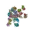
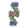
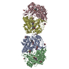
| Title | Crystal Structure of Prefusion-stabilized RSV F variant SC-TM | ||||||
 Components Components | Fusion glycoprotein F0,Fibritin | ||||||
 Keywords Keywords |  VIRAL PROTEIN / class I viral fusion protein / fusion / respiratory syncytial virus / prefusion VIRAL PROTEIN / class I viral fusion protein / fusion / respiratory syncytial virus / prefusion | ||||||
| Function / homology |  Function and homology information Function and homology informationpositive regulation of syncytium formation by virus / host cell Golgi membrane / entry receptor-mediated virion attachment to host cell / symbiont entry into host cell / fusion of virus membrane with host plasma membrane / viral envelope / host cell plasma membrane / virion membrane / membrane / identical protein bindingSimilarity search - Function | ||||||
| Biological species |  Human respiratory syncytial virus AEnterobacteria phage Ox2 (virus) Human respiratory syncytial virus AEnterobacteria phage Ox2 (virus) | ||||||
| Method | X-RAY DIFFRACTION / SYNCHROTRON / MOLECULAR REPLACEMENT / molecular replacement / Resolution: 2.4 Å | ||||||
 Authors Authors | McLellan, J.S. / Langedijk, J.P.M. | ||||||
 Citation Citation | Journal: Nat Commun / Year: 2015 Title: A highly stable prefusion RSV F vaccine derived from structural analysis of the fusion mechanism. Authors: Krarup, A. / Truan, D. / Furmanova-Hollenstein, P. / Bogaert, L. / Bouchier, P. / Bisschop, I.J. / Widjojoatmodjo, M.N. / Zahn, R. / Schuitemaker, H. / McLellan, J.S. / Langedijk, J.P. | ||||||
| History |
|
- Structure visualization
| Structure viewer | Molecule: MolmilJmol/JSmol |
|---|
- Downloads & links
-Download
| PDBx/mmCIF format | 5c6b.cif.gz | 201 KB | Display | PDBx/mmCIF format |
|---|---|---|---|---|
| PDB format | pdb5c6b.ent.gz | 157 KB | Display | PDB format |
| PDBx/mmJSON format | 5c6b.json.gz | Tree view | PDBx/mmJSON format | |
| Others |  Other downloads Other downloads |
-Validation report
| Arichive directory | https://data.pdbj.org/pub/pdb/validation_reports/c6/5c6bftp://data.pdbj.org/pub/pdb/validation_reports/c6/5c6b | HTTPS FTP |
|---|
-Related structure data
| Related structure data |  5c69C  4mmsS C: citing same article ( S: Starting model for refinement |
|---|---|
| Similar structure data |


-Links
 PDBj
PDBj


- Assembly
| Deposited unit | 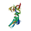
| ||||||||||||||||||||||||
|---|---|---|---|---|---|---|---|---|---|---|---|---|---|---|---|---|---|---|---|---|---|---|---|---|---|
| 1 | x 6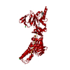
| ||||||||||||||||||||||||
| Unit cell |
| ||||||||||||||||||||||||
| Components on special symmetry positions |
|
-Components
| #1: Protein | Mass: 54712.277 Da / Num. of mol.: 1 / Fragment: ectodomain Mutation: N67I, S215P, E487Q, I379V, M447V,N67I, S215P, E487Q, I379V, M447V Source method: isolated from a genetically manipulated source Source: (gene. exp.) Human respiratory syncytial virus A, (gene. exp.) Enterobacteria phage Ox2 (virus)Strain: A2 / Gene: wac / Cell line (production host): HEK293 / Production host:  homo sapiens (human) / References: UniProt: P03420, UniProt: Q38650 homo sapiens (human) / References: UniProt: P03420, UniProt: Q38650 | ||||
|---|---|---|---|---|---|
| #2: Chemical | ChemComp-NHE / CHES (buffer)  Mass: 207.290 Da / Num. of mol.: 1 / Source method: obtained synthetically / Formula: C8H17NO3S / Comment: pH buffer*YM Mass: 207.290 Da / Num. of mol.: 1 / Source method: obtained synthetically / Formula: C8H17NO3S / Comment: pH buffer*YM | ||||
| #3: Chemical | ChemComp-SO4 / Sulfate  Mass: 96.063 Da / Num. of mol.: 4 / Source method: obtained synthetically / Formula: SO4 Mass: 96.063 Da / Num. of mol.: 4 / Source method: obtained synthetically / Formula: SO4#4: Chemical | Chloride  Mass: 35.453 Da / Num. of mol.: 2 / Source method: obtained synthetically / Formula: Cl Mass: 35.453 Da / Num. of mol.: 2 / Source method: obtained synthetically / Formula: Cl#5: Water | ChemComp-HOH / | Water Mass: 18.015 Da / Num. of mol.: 161 / Source method: isolated from a natural source / Formula: H2O Mass: 18.015 Da / Num. of mol.: 161 / Source method: isolated from a natural source / Formula: H2O |
-Experimental details
-Experiment
| Experiment | Method: X-RAY DIFFRACTION / Number of used crystals: 1 |
|---|
- Sample preparation
| Crystal | Density Matthews: 3.62 Å3/Da / Density % sol: 66.06 % |
|---|---|
| Crystal grow | Temperature: 293 K / Method: vapor diffusion / pH: 9.5 / Details: 1.34M K/Na tartrate, 0.2M LiSO4, 0.1M CHES pH 9.5 |
-Data collection
| Diffraction | Mean temperature: 100 K |
|---|---|
| Diffraction source | Source: SYNCHROTRON / Site: APS  / Beamline: 19-ID / Wavelength: 0.9792368 Å / Beamline: 19-ID / Wavelength: 0.9792368 Å |
| Detector | Type: ADSC QUANTUM 315 / Detector: CCD / Date: Nov 11, 2014 |
| Radiation | Protocol: SINGLE WAVELENGTH / Monochromatic (M) / Laue (L): M / Scattering type: x-ray |
| Radiation wavelength | Wavelength: 0.9792368 Å / Relative weight: 1 |
| Reflection | Resolution: 2.4→44.95 Å / Num. all: 32205 / Num. obs: 32205 / % possible obs: 99.7 % / Redundancy: 10.5 % / Net I/σ(I): 12.6 |
| Reflection shell | Resolution: 2.4→2.49 Å / Redundancy: 10.7 % / Rmerge(I) obs: 1.327 / Mean I/σ(I) obs: 1.9 / % possible all: 100 |
-Phasing
| Phasing | Method: molecular replacement |
|---|
- Processing
| Software |
| |||||||||||||||||||||||||||||||||||||||||||||||||||||||||||||||||||||||||||||||||||||||||||||||||||||||||||||||||||||||||||||||||||||||||||||||||||||||||||||||||||||||||||||||
|---|---|---|---|---|---|---|---|---|---|---|---|---|---|---|---|---|---|---|---|---|---|---|---|---|---|---|---|---|---|---|---|---|---|---|---|---|---|---|---|---|---|---|---|---|---|---|---|---|---|---|---|---|---|---|---|---|---|---|---|---|---|---|---|---|---|---|---|---|---|---|---|---|---|---|---|---|---|---|---|---|---|---|---|---|---|---|---|---|---|---|---|---|---|---|---|---|---|---|---|---|---|---|---|---|---|---|---|---|---|---|---|---|---|---|---|---|---|---|---|---|---|---|---|---|---|---|---|---|---|---|---|---|---|---|---|---|---|---|---|---|---|---|---|---|---|---|---|---|---|---|---|---|---|---|---|---|---|---|---|---|---|---|---|---|---|---|---|---|---|---|---|---|---|---|---|---|
| Refinement | Method to determine structure: MOLECULAR REPLACEMENT Starting model: 4MMS Resolution: 2.4→44.95 Å / SU ML: 0.3 / Cross valid method: FREE R-VALUE / σ(F): 1.34 / Phase error: 22.2 / Stereochemistry target values: ML
| |||||||||||||||||||||||||||||||||||||||||||||||||||||||||||||||||||||||||||||||||||||||||||||||||||||||||||||||||||||||||||||||||||||||||||||||||||||||||||||||||||||||||||||||
| Solvent computation | Shrinkage radii: 0.9 Å / VDW probe radii: 1.11 Å / Solvent model: FLAT BULK SOLVENT MODEL | |||||||||||||||||||||||||||||||||||||||||||||||||||||||||||||||||||||||||||||||||||||||||||||||||||||||||||||||||||||||||||||||||||||||||||||||||||||||||||||||||||||||||||||||
| Displacement parameters | Biso max: 176.28 Å2 / Biso mean: 56.316 Å2 / Biso min: 19.77 Å2 | |||||||||||||||||||||||||||||||||||||||||||||||||||||||||||||||||||||||||||||||||||||||||||||||||||||||||||||||||||||||||||||||||||||||||||||||||||||||||||||||||||||||||||||||
| Refinement step | Cycle: final / Resolution: 2.4→44.95 Å
| |||||||||||||||||||||||||||||||||||||||||||||||||||||||||||||||||||||||||||||||||||||||||||||||||||||||||||||||||||||||||||||||||||||||||||||||||||||||||||||||||||||||||||||||
| Refine LS restraints |
| |||||||||||||||||||||||||||||||||||||||||||||||||||||||||||||||||||||||||||||||||||||||||||||||||||||||||||||||||||||||||||||||||||||||||||||||||||||||||||||||||||||||||||||||
| LS refinement shell | Refine-ID: X-RAY DIFFRACTION / Total num. of bins used: 12
| |||||||||||||||||||||||||||||||||||||||||||||||||||||||||||||||||||||||||||||||||||||||||||||||||||||||||||||||||||||||||||||||||||||||||||||||||||||||||||||||||||||||||||||||
| Refinement TLS params. | Method: refined / Refine-ID: X-RAY DIFFRACTION
| |||||||||||||||||||||||||||||||||||||||||||||||||||||||||||||||||||||||||||||||||||||||||||||||||||||||||||||||||||||||||||||||||||||||||||||||||||||||||||||||||||||||||||||||
| Refinement TLS group |
|
