 Movie
Movie Controller

[English] 日本語
 Yorodumi
- PDB-4zyp: Crystal Structure of Motavizumab and Quaternary-Specific RSV-Neut... -
+ Open data

- Basic information
| Entry | Database: PDB / ID: 4zyp | ||||||
|---|---|---|---|---|---|---|---|
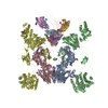
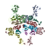
| Title | Crystal Structure of Motavizumab and Quaternary-Specific RSV-Neutralizing Human Antibody AM14 in Complex with Prefusion RSV F Glycoprotein | ||||||
 Components Components |
| ||||||
 Keywords Keywords |  IMMUNE SYSTEM / Ig domain / Fab / fusion / respiratory syncytial virus / prefusion IMMUNE SYSTEM / Ig domain / Fab / fusion / respiratory syncytial virus / prefusion | ||||||
| Function / homology |  Function and homology information Function and homology informationpositive regulation of syncytium formation by virus / host cell Golgi membrane / entry receptor-mediated virion attachment to host cell / symbiont entry into host cell / fusion of virus membrane with host plasma membrane / viral envelope / host cell plasma membrane / virion membrane / membrane / identical protein bindingSimilarity search - Function | ||||||
| Biological species |  Human respiratory syncytial virus AEnterobacteria phage T4 (virus) Human respiratory syncytial virus AEnterobacteria phage T4 (virus) Mus musculus (house mouse) Mus musculus (house mouse) Homo sapiens (human) Homo sapiens (human) | ||||||
| Method | X-RAY DIFFRACTION / SYNCHROTRON / MOLECULAR REPLACEMENT / Resolution: 5.5 Å | ||||||
 Authors Authors | Gilman, M.S.A. / McLellan, J.S. | ||||||
 Citation Citation | Journal: Plos Pathog. / Year: 2015 Title: Characterization of a Prefusion-Specific Antibody That Recognizes a Quaternary, Cleavage-Dependent Epitope on the RSV Fusion Glycoprotein. Authors: Gilman, M.S. / Moin, S.M. / Mas, V. / Chen, M. / Patel, N.K. / Kramer, K. / Zhu, Q. / Kabeche, S.C. / Kumar, A. / Palomo, C. / Beaumont, T. / Baxa, U. / Ulbrandt, N.D. / Melero, J.A. / ...Authors: Gilman, M.S. / Moin, S.M. / Mas, V. / Chen, M. / Patel, N.K. / Kramer, K. / Zhu, Q. / Kabeche, S.C. / Kumar, A. / Palomo, C. / Beaumont, T. / Baxa, U. / Ulbrandt, N.D. / Melero, J.A. / Graham, B.S. / McLellan, J.S. | ||||||
| History |
|
- Structure visualization
| Structure viewer | Molecule: MolmilJmol/JSmol |
|---|
- Downloads & links
-Download
| PDBx/mmCIF format | 4zyp.cif.gz | 678.9 KB | Display | PDBx/mmCIF format |
|---|---|---|---|---|
| PDB format | pdb4zyp.ent.gz | 565.1 KB | Display | PDB format |
| PDBx/mmJSON format | 4zyp.json.gz | Tree view | PDBx/mmJSON format | |
| Others |  Other downloads Other downloads |
-Validation report
| Arichive directory | https://data.pdbj.org/pub/pdb/validation_reports/zy/4zypftp://data.pdbj.org/pub/pdb/validation_reports/zy/4zyp | HTTPS FTP |
|---|
-Related structure data
| Related structure data | 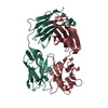 4zykSC  3ixtS  4jhwS S: Starting model for refinement C: citing same article ( |
|---|---|
| Similar structure data |


-Links
 PDBj
PDBj


- Assembly
| Deposited unit | 
| ||||||||
|---|---|---|---|---|---|---|---|---|---|
| 1 |
| ||||||||
| Unit cell |
|
-Components
| #1: Protein | Mass: 55120.879 Da / Num. of mol.: 3 / Mutation: S155C, S190F, V207L, S290C, I379V, M447V Source method: isolated from a genetically manipulated source Source: (gene. exp.) Human respiratory syncytial virus A (strain A2), (gene. exp.) Enterobacteria phage T4 (virus)Strain: A2 / Plasmid: p(alpha)H / Gene: wac, T4Tp161 / Cell line (production host): HEK293F / Production host: Homo sapiens (human) / References: UniProt: P03420, UniProt: D9IEJ2#2: Antibody | Mass: 24284.465 Da / Num. of mol.: 3 Source method: isolated from a genetically manipulated source Details: Humanized mouse antibody / Source: (gene. exp.) Mus musculus (house mouse) / Plasmid: pVRC8400 / Cell line (production host): HEK293F / Production host: Homo sapiens (human)#3: Antibody | Mass: 23150.730 Da / Num. of mol.: 3 Source method: isolated from a genetically manipulated source Details: Humanized mouse antibody / Source: (gene. exp.) Mus musculus (house mouse) / Plasmid: pVRC8400 / Production host: Homo sapiens (human)#4: Antibody | Mass: 24365.150 Da / Num. of mol.: 3 Source method: isolated from a genetically manipulated source Source: (gene. exp.) Homo sapiens (human) / Plasmid: pVRC8400 / Cell line (production host): HEK293F / Production host: Homo sapiens (human)#5: Antibody | Mass: 23615.227 Da / Num. of mol.: 3 Source method: isolated from a genetically manipulated source Source: (gene. exp.) Homo sapiens (human) / Plasmid: pVRC8400 / Cell line (production host): HEK293F / Production host: Homo sapiens (human) |
|---|
-Experimental details
-Experiment
| Experiment | Method: X-RAY DIFFRACTION / Number of used crystals: 1 |
|---|
- Sample preparation
| Crystal | Density Matthews: 3.1 Å3/Da / Density % sol: 60.26 % |
|---|---|
| Crystal grow | Temperature: 293 K / Method: vapor diffusion, hanging drop / pH: 6.5 Details: 5.645mg/mL EndoH digested DS-Cav1 + AM14 Fab + Motavizumab Fab, 11.4% PEG8000, 1.71% MPD, 0.1M Imidazole pH 6.5 |
-Data collection
| Diffraction | Mean temperature: 100 K | |||||||||||||||||||||||||||
|---|---|---|---|---|---|---|---|---|---|---|---|---|---|---|---|---|---|---|---|---|---|---|---|---|---|---|---|---|
| Diffraction source | Source: SYNCHROTRON / Site: APS  / Beamline: 19-ID / Wavelength: 0.9792 Å / Beamline: 19-ID / Wavelength: 0.9792 Å | |||||||||||||||||||||||||||
| Detector | Type: ADSC QUANTUM 315r / Detector: CCD / Date: Jul 20, 2014 | |||||||||||||||||||||||||||
| Radiation | Protocol: SINGLE WAVELENGTH / Monochromatic (M) / Laue (L): M / Scattering type: x-ray | |||||||||||||||||||||||||||
| Radiation wavelength | Wavelength: 0.9792 Å / Relative weight: 1 | |||||||||||||||||||||||||||
| Reflection | Resolution: 5.5→49.59 Å / Num. obs: 17434 / % possible obs: 97.5 % / Redundancy: 3 % / Biso Wilson estimate: 161.85 Å2 / CC1/2: 0.939 / Rmerge(I) obs: 0.182 / Rpim(I) all: 0.125 / Net I/σ(I): 4.8 / Num. measured all: 51879 / Scaling rejects: 33 | |||||||||||||||||||||||||||
| Reflection shell | Diffraction-ID: 1 / Rejects: 0
|
- Processing
| Software |
| |||||||||||||||||||||||||||||||||||||||||||||||||
|---|---|---|---|---|---|---|---|---|---|---|---|---|---|---|---|---|---|---|---|---|---|---|---|---|---|---|---|---|---|---|---|---|---|---|---|---|---|---|---|---|---|---|---|---|---|---|---|---|---|---|
| Refinement | Method to determine structure: MOLECULAR REPLACEMENT Starting model: 4JHW, 3IXT, 4ZYK Resolution: 5.5→49.386 Å / SU ML: 0.91 / Cross valid method: FREE R-VALUE / σ(F): 1.35 / Phase error: 32.56 / Stereochemistry target values: ML
| |||||||||||||||||||||||||||||||||||||||||||||||||
| Solvent computation | Shrinkage radii: 0.9 Å / VDW probe radii: 1.11 Å / Solvent model: FLAT BULK SOLVENT MODEL | |||||||||||||||||||||||||||||||||||||||||||||||||
| Refinement step | Cycle: LAST / Resolution: 5.5→49.386 Å
| |||||||||||||||||||||||||||||||||||||||||||||||||
| Refine LS restraints |
| |||||||||||||||||||||||||||||||||||||||||||||||||
| LS refinement shell |
|
