 Movie
Movie Controller

[English] 日本語
 Yorodumi
- PDB-5tfw: Crystal structure of 10E8 Fab light chain mutant2 against the MPE... -
+ Open data

- Basic information
| Entry | Database: PDB / ID: 5tfw | |||||||||
|---|---|---|---|---|---|---|---|---|---|---|
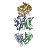
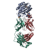
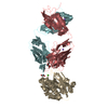
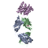
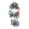
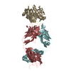
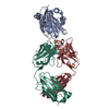
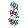
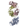
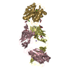
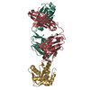
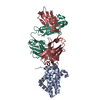
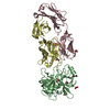
| Title | Crystal structure of 10E8 Fab light chain mutant2 against the MPER region of the HIV-1 Env, in complex with T117v2 epitope scaffold | |||||||||
 Components Components |
| |||||||||
 Keywords Keywords |  IMMUNE SYSTEM / HIV-1 GP41 MPER / 10E8 FAB / LIPID MEMBRANE IMMUNE SYSTEM / HIV-1 GP41 MPER / 10E8 FAB / LIPID MEMBRANE | |||||||||
| Function / homology | Mannitol-specific EII; Chain A / Mannitol-specific EII; Chain A / Immunoglobulins / Immunoglobulin-like / Sandwich / 3-Layer(aba) Sandwich / Mainly Beta / Alpha Beta / DI(HYDROXYETHYL)ETHER Function and homology information Function and homology information | |||||||||
| Biological species |  Homo sapiens (human) Homo sapiens (human)synthetic construct (others) | |||||||||
| Method | X-RAY DIFFRACTION / SYNCHROTRON / MOLECULAR REPLACEMENT / Resolution: 2.168 Å | |||||||||
 Authors Authors | Irimia, A. / Wilson, I.A. | |||||||||
| Funding support |  United States, 2items United States, 2items
| |||||||||
 Citation Citation | Journal: PLoS Pathog. / Year: 2017 Title: Lipid interactions and angle of approach to the HIV-1 viral membrane of broadly neutralizing antibody 10E8: Insights for vaccine and therapeutic design. Authors: Irimia, A. / Serra, A.M. / Sarkar, A. / Jacak, R. / Kalyuzhniy, O. / Sok, D. / Saye-Francisco, K.L. / Schiffner, T. / Tingle, R. / Kubitz, M. / Adachi, Y. / Stanfield, R.L. / Deller, M.C. / ...Authors: Irimia, A. / Serra, A.M. / Sarkar, A. / Jacak, R. / Kalyuzhniy, O. / Sok, D. / Saye-Francisco, K.L. / Schiffner, T. / Tingle, R. / Kubitz, M. / Adachi, Y. / Stanfield, R.L. / Deller, M.C. / Burton, D.R. / Schief, W.R. / Wilson, I.A. | |||||||||
| History |
|
- Structure visualization
| Structure viewer | Molecule: MolmilJmol/JSmol |
|---|
- Downloads & links
-Download
| PDBx/mmCIF format | 5tfw.cif.gz | 253.2 KB | Display | PDBx/mmCIF format |
|---|---|---|---|---|
| PDB format | pdb5tfw.ent.gz | 202.5 KB | Display | PDB format |
| PDBx/mmJSON format | 5tfw.json.gz | Tree view | PDBx/mmJSON format | |
| Others |  Other downloads Other downloads |
-Validation report
| Arichive directory | https://data.pdbj.org/pub/pdb/validation_reports/tf/5tfwftp://data.pdbj.org/pub/pdb/validation_reports/tf/5tfw | HTTPS FTP |
|---|
-Related structure data
| Related structure data |  5sy8C  5t29C  5t5bC  5t6lC  5t80C  5t85C  3lf6S  4g6fS S: Starting model for refinement C: citing same article ( |
|---|---|
| Similar structure data |


-Links
 PDBj
PDBj

- Assembly
| Deposited unit | 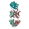
| ||||||||
|---|---|---|---|---|---|---|---|---|---|
| 1 |
| ||||||||
| Unit cell |
|
-Components
-Protein , 1 types, 1 molecules O
| #3: Protein | Mass: 18643.090 Da / Num. of mol.: 1 Source method: isolated from a genetically manipulated source Source: (gene. exp.) synthetic construct (others) / Plasmid: PET29 / Production host:  Escherichia coli (E. coli) / Strain (production host): BL-21 STAR Escherichia coli (E. coli) / Strain (production host): BL-21 STAR |
|---|
-Antibody , 2 types, 2 molecules HL
| #1: Antibody | Mass: 25447.475 Da / Num. of mol.: 1 Source method: isolated from a genetically manipulated source Source: (gene. exp.) Homo sapiens (human) / Gene: IGHV3-15*05 / Plasmid: PHCMV3 / Cell line (production host): HEK 293S / Production host: Homo sapiens (human) |
|---|---|
| #2: Antibody | Mass: 22863.111 Da / Num. of mol.: 1 Source method: isolated from a genetically manipulated source Source: (gene. exp.) Homo sapiens (human) / Gene: IGLV3-19*01 / Plasmid: PHCMV3 / Cell line (production host): HEK 293S / Production host: Homo sapiens (human) |
-Non-polymers , 3 types, 233 molecules 


| #4: Chemical | ChemComp-PEG / Diethylene glycol Mass: 106.120 Da / Num. of mol.: 1 / Source method: obtained synthetically / Formula: C4H10O3 Mass: 106.120 Da / Num. of mol.: 1 / Source method: obtained synthetically / Formula: C4H10O3 |
|---|---|
| #5: Chemical | ChemComp-EDO / Ethylene glycol Mass: 62.068 Da / Num. of mol.: 1 / Source method: obtained synthetically / Formula: C2H6O2 Mass: 62.068 Da / Num. of mol.: 1 / Source method: obtained synthetically / Formula: C2H6O2 |
| #6: Water | ChemComp-HOH / WaterMass: 18.015 Da / Num. of mol.: 231 / Source method: isolated from a natural source / Formula: H2O |
-Experimental details
-Experiment
| Experiment | Method: X-RAY DIFFRACTION / Number of used crystals: 1 |
|---|
- Sample preparation
| Crystal | Density Matthews: 2.8 Å3/Da / Density % sol: 55.8 % |
|---|---|
| Crystal grow | Temperature: 295.15 K / Method: vapor diffusion / pH: 6.9 Details: 0.2 M Na thiocyanate, 20% PEG 3350, pH 6.9, 10% ethylene glycol |
-Data collection
| Diffraction | Mean temperature: 110 K |
|---|---|
| Diffraction source | Source: SYNCHROTRON / Site: APS / Beamline: 23-ID-B / Wavelength: 1.0332 Å |
| Detector | Type: MARMOSAIC 300 mm CCD / Detector: CCD / Date: Jul 25, 2015 |
| Radiation | Protocol: SINGLE WAVELENGTH / Monochromatic (M) / Laue (L): M / Scattering type: x-ray |
| Radiation wavelength | Wavelength: 1.0332 Å / Relative weight: 1 |
| Reflection | Resolution: 2.168→47.235 Å / Num. obs: 36138 / % possible obs: 94.5 % / Redundancy: 4.2 % / Biso Wilson estimate: 41 Å2 / CC1/2: 0.999 / Rsym value: 0.056 / Net I/σ(I): 15.5 |
| Reflection shell | Resolution: 2.168→2.22 Å / Redundancy: 4 % / Rmerge(I) obs: 0.635 / Mean I/σ(I) obs: 2.1 / CC1/2: 0.835 / % possible all: 95.8 |
- Processing
| Software |
| ||||||||||||||||||||||||||||||||||||||||||||||||||||||||||||||||||||||||||||||||||||||||||||||||||||||||||||||||||||||||||||||||||||||||||||||||||||||
|---|---|---|---|---|---|---|---|---|---|---|---|---|---|---|---|---|---|---|---|---|---|---|---|---|---|---|---|---|---|---|---|---|---|---|---|---|---|---|---|---|---|---|---|---|---|---|---|---|---|---|---|---|---|---|---|---|---|---|---|---|---|---|---|---|---|---|---|---|---|---|---|---|---|---|---|---|---|---|---|---|---|---|---|---|---|---|---|---|---|---|---|---|---|---|---|---|---|---|---|---|---|---|---|---|---|---|---|---|---|---|---|---|---|---|---|---|---|---|---|---|---|---|---|---|---|---|---|---|---|---|---|---|---|---|---|---|---|---|---|---|---|---|---|---|---|---|---|---|---|---|---|
| Refinement | Method to determine structure: MOLECULAR REPLACEMENT Starting model: 4G6F, 3LF6 Resolution: 2.168→47.235 Å / SU ML: 0.27 / Cross valid method: FREE R-VALUE / σ(F): 1.35 / Phase error: 26.77 / Stereochemistry target values: ML
| ||||||||||||||||||||||||||||||||||||||||||||||||||||||||||||||||||||||||||||||||||||||||||||||||||||||||||||||||||||||||||||||||||||||||||||||||||||||
| Solvent computation | Shrinkage radii: 0.9 Å / VDW probe radii: 1.11 Å / Solvent model: FLAT BULK SOLVENT MODEL | ||||||||||||||||||||||||||||||||||||||||||||||||||||||||||||||||||||||||||||||||||||||||||||||||||||||||||||||||||||||||||||||||||||||||||||||||||||||
| Displacement parameters | Biso mean: 59 Å2 | ||||||||||||||||||||||||||||||||||||||||||||||||||||||||||||||||||||||||||||||||||||||||||||||||||||||||||||||||||||||||||||||||||||||||||||||||||||||
| Refinement step | Cycle: LAST / Resolution: 2.168→47.235 Å
| ||||||||||||||||||||||||||||||||||||||||||||||||||||||||||||||||||||||||||||||||||||||||||||||||||||||||||||||||||||||||||||||||||||||||||||||||||||||
| Refine LS restraints |
| ||||||||||||||||||||||||||||||||||||||||||||||||||||||||||||||||||||||||||||||||||||||||||||||||||||||||||||||||||||||||||||||||||||||||||||||||||||||
| LS refinement shell |
| ||||||||||||||||||||||||||||||||||||||||||||||||||||||||||||||||||||||||||||||||||||||||||||||||||||||||||||||||||||||||||||||||||||||||||||||||||||||
| Refinement TLS params. | Method: refined / Refine-ID: X-RAY DIFFRACTION
| ||||||||||||||||||||||||||||||||||||||||||||||||||||||||||||||||||||||||||||||||||||||||||||||||||||||||||||||||||||||||||||||||||||||||||||||||||||||
| Refinement TLS group |
|
