 Movie
Movie Controller


+ Open data

- Basic information
| Entry | Database: PDB / ID: 5te3 | |||||||||
|---|---|---|---|---|---|---|---|---|---|---|
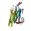
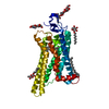
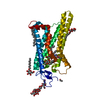
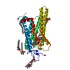
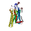
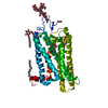
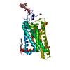
| Title | Crystal structure of Bos taurus opsin at 2.7 Angstrom | |||||||||
 Components Components | Rhodopsin | |||||||||
 Keywords Keywords | SIGNALING PROTEIN | |||||||||
| Function / homology |  Function and homology informationOpsins / VxPx cargo-targeting to cilium / rod bipolar cell differentiation / rod photoreceptor outer segment / sperm head plasma membrane / podosome assembly / absorption of visible light / opsin binding / The canonical retinoid cycle in rods (twilight vision) / : ...Opsins / VxPx cargo-targeting to cilium / rod bipolar cell differentiation / rod photoreceptor outer segment / sperm head plasma membrane / podosome assembly / absorption of visible light / opsin binding / The canonical retinoid cycle in rods (twilight vision) / : / G protein-coupled photoreceptor activity / photoreceptor inner segment membrane / G protein-coupled opsin signaling pathway / 11-cis retinal binding / cellular response to light stimulus / G protein-coupled receptor complex / Inactivation, recovery and regulation of the phototransduction cascade / phototransduction, visible light / thermotaxis / Activation of the phototransduction cascade / outer membrane / detection of temperature stimulus involved in thermoception / arrestin family protein binding / photoreceptor cell maintenance / photoreceptor outer segment membrane / G alpha (i) signalling events / phototransduction / response to light stimulus / photoreceptor outer segment / G-protein alpha-subunit binding / sperm midpiece / visual perception / guanyl-nucleotide exchange factor activity / microtubule cytoskeleton organization / photoreceptor disc membrane / cell-cell junction / gene expression / G protein-coupled receptor signaling pathway / Golgi membrane / zinc ion binding / membrane / identical protein binding / plasma membrane Function and homology informationOpsins / VxPx cargo-targeting to cilium / rod bipolar cell differentiation / rod photoreceptor outer segment / sperm head plasma membrane / podosome assembly / absorption of visible light / opsin binding / The canonical retinoid cycle in rods (twilight vision) / : ...Opsins / VxPx cargo-targeting to cilium / rod bipolar cell differentiation / rod photoreceptor outer segment / sperm head plasma membrane / podosome assembly / absorption of visible light / opsin binding / The canonical retinoid cycle in rods (twilight vision) / : / G protein-coupled photoreceptor activity / photoreceptor inner segment membrane / G protein-coupled opsin signaling pathway / 11-cis retinal binding / cellular response to light stimulus / G protein-coupled receptor complex / Inactivation, recovery and regulation of the phototransduction cascade / phototransduction, visible light / thermotaxis / Activation of the phototransduction cascade / outer membrane / detection of temperature stimulus involved in thermoception / arrestin family protein binding / photoreceptor cell maintenance / photoreceptor outer segment membrane / G alpha (i) signalling events / phototransduction / response to light stimulus / photoreceptor outer segment / G-protein alpha-subunit binding / sperm midpiece / visual perception / guanyl-nucleotide exchange factor activity / microtubule cytoskeleton organization / photoreceptor disc membrane / cell-cell junction / gene expression / G protein-coupled receptor signaling pathway / Golgi membrane / zinc ion binding / membrane / identical protein binding / plasma membraneSimilarity search - Function | |||||||||
| Biological species |  Bos taurus (cattle) Bos taurus (cattle) | |||||||||
| Method | X-RAY DIFFRACTION / SYNCHROTRON / FOURIER SYNTHESIS / Resolution: 2.7 Å | |||||||||
 Authors Authors | Gulati, S. / Kiser, P.D. / Palczewski, K. | |||||||||
 Citation Citation | Journal: Proc. Natl. Acad. Sci. U.S.A. / Year: 2017 Title: Photocyclic behavior of rhodopsin induced by an atypical isomerization mechanism. Authors: Gulati, S. / Jastrzebska, B. / Banerjee, S. / Placeres, A.L. / Miszta, P. / Gao, S. / Gunderson, K. / Tochtrop, G.P. / Filipek, S. / Katayama, K. / Kiser, P.D. / Mogi, M. / Stewart, P.L. / Palczewski, K. | |||||||||
| History |
|
- Structure visualization
| Structure viewer | Molecule: MolmilJmol/JSmol |
|---|
- Downloads & links
-Download
| PDBx/mmCIF format | 5te3.cif.gz | 86.3 KB | Display | PDBx/mmCIF format |
|---|---|---|---|---|
| PDB format | pdb5te3.ent.gz | 62.6 KB | Display | PDB format |
| PDBx/mmJSON format | 5te3.json.gz | Tree view | PDBx/mmJSON format | |
| Others |  Other downloads Other downloads |
-Validation report
| Arichive directory | https://data.pdbj.org/pub/pdb/validation_reports/te/5te3ftp://data.pdbj.org/pub/pdb/validation_reports/te/5te3 | HTTPS FTP |
|---|
-Related structure data
| Related structure data |  5te5C 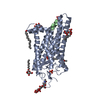 4j4qS C: citing same article ( S: Starting model for refinement |
|---|---|
| Similar structure data |


-Links
 PDBj
PDBj


- Assembly
| Deposited unit | 
| ||||||||
|---|---|---|---|---|---|---|---|---|---|
| 1 |
| ||||||||
| 2 | 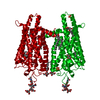
| ||||||||
| Unit cell |
| ||||||||
| Components on special symmetry positions |
|
-Components
-Protein , 1 types, 1 molecules A
| #1: Protein | Mass: 39031.457 Da / Num. of mol.: 1 / Source method: isolated from a natural source / Source: (natural) Bos taurus (cattle) / References: UniProt: P02699 |
|---|
-Sugars , 2 types, 4 molecules 
| #2: Polysaccharide | alpha-D-mannopyranose-(1-3)-beta-D-mannopyranose-(1-4)-2-acetamido-2-deoxy-beta-D-glucopyranose-(1- ...alpha-D-mannopyranose-(1-3)-beta-D-mannopyranose-(1-4)-2-acetamido-2-deoxy-beta-D-glucopyranose-(1-4)-2-acetamido-2-deoxy-beta-D-glucopyranose / Mass: 748.682 Da / Num. of mol.: 1 Source method: isolated from a genetically manipulated source |
|---|---|
| #3: Sugar | Octyl glucoside Type: D-saccharide / Mass: 292.369 Da / Num. of mol.: 3 Type: D-saccharide / Mass: 292.369 Da / Num. of mol.: 3Source method: isolated from a genetically manipulated source Formula: C14H28O6 / Comment: detergent*YM |
-Non-polymers , 3 types, 36 molecules 


| #4: Chemical | Palmitic acid Mass: 256.424 Da / Num. of mol.: 2 / Source method: obtained synthetically / Formula: C16H32O2 Mass: 256.424 Da / Num. of mol.: 2 / Source method: obtained synthetically / Formula: C16H32O2#5: Chemical | Sulfate Mass: 96.063 Da / Num. of mol.: 2 / Source method: obtained synthetically / Formula: SO4 Mass: 96.063 Da / Num. of mol.: 2 / Source method: obtained synthetically / Formula: SO4#6: Water | ChemComp-HOH / | WaterMass: 18.015 Da / Num. of mol.: 32 / Source method: isolated from a natural source / Formula: H2O |
|---|
-Experimental details
-Experiment
| Experiment | Method: X-RAY DIFFRACTION / Number of used crystals: 1 |
|---|
- Sample preparation
| Crystal | Density % sol: 84.84 % |
|---|---|
| Crystal grow | Temperature: 277 K / Method: vapor diffusion, hanging drop Details: contained 2.8-3.4 M ammonium sulfate in 0.05-0.1 M NaAcO buffer, pH 5.2-5.6 PH range: 5.2-5.6 |
-Data collection
| Diffraction | Mean temperature: 100 K |
|---|---|
| Diffraction source | Source: SYNCHROTRON / Site: APS  / Beamline: 24-ID-C / Wavelength: 0.9789 Å / Beamline: 24-ID-C / Wavelength: 0.9789 Å |
| Detector | Type: DECTRIS PILATUS 6M-F / Detector: PIXEL / Date: Feb 27, 2014 |
| Radiation | Protocol: SINGLE WAVELENGTH / Monochromatic (M) / Laue (L): M / Scattering type: x-ray |
| Radiation wavelength | Wavelength: 0.9789 Å / Relative weight: 1 |
| Reflection | Resolution: 2.7→50 Å / Num. obs: 34545 / % possible obs: 100 % / Redundancy: 22.8 % / Net I/σ(I): 20.9 |
- Processing
| Software |
| ||||||||||||||||||||||||||||||||||||||||||||||||||||||||||||||||||||||||||||||||||||||||||||||||||||||||||||||||||||||||||||||||||||||||||||||||||||||||||||||||||||||||||||||||||||||
|---|---|---|---|---|---|---|---|---|---|---|---|---|---|---|---|---|---|---|---|---|---|---|---|---|---|---|---|---|---|---|---|---|---|---|---|---|---|---|---|---|---|---|---|---|---|---|---|---|---|---|---|---|---|---|---|---|---|---|---|---|---|---|---|---|---|---|---|---|---|---|---|---|---|---|---|---|---|---|---|---|---|---|---|---|---|---|---|---|---|---|---|---|---|---|---|---|---|---|---|---|---|---|---|---|---|---|---|---|---|---|---|---|---|---|---|---|---|---|---|---|---|---|---|---|---|---|---|---|---|---|---|---|---|---|---|---|---|---|---|---|---|---|---|---|---|---|---|---|---|---|---|---|---|---|---|---|---|---|---|---|---|---|---|---|---|---|---|---|---|---|---|---|---|---|---|---|---|---|---|---|---|---|---|
| Refinement | Method to determine structure: FOURIER SYNTHESIS Starting model: 4J4Q Resolution: 2.7→49.34 Å / Cor.coef. Fo:Fc: 0.927 / Cor.coef. Fo:Fc free: 0.939 / SU B: 12.971 / SU ML: 0.223 / Cross valid method: THROUGHOUT / ESU R: 0.242 / ESU R Free: 0.203 / Stereochemistry target values: MAXIMUM LIKELIHOOD / Details: HYDROGENS HAVE BEEN ADDED IN THE RIDING POSITIONS
| ||||||||||||||||||||||||||||||||||||||||||||||||||||||||||||||||||||||||||||||||||||||||||||||||||||||||||||||||||||||||||||||||||||||||||||||||||||||||||||||||||||||||||||||||||||||
| Solvent computation | Ion probe radii: 0.8 Å / Shrinkage radii: 0.8 Å / VDW probe radii: 1.2 Å / Solvent model: MASK | ||||||||||||||||||||||||||||||||||||||||||||||||||||||||||||||||||||||||||||||||||||||||||||||||||||||||||||||||||||||||||||||||||||||||||||||||||||||||||||||||||||||||||||||||||||||
| Displacement parameters | Biso mean: 94.944 Å2
| ||||||||||||||||||||||||||||||||||||||||||||||||||||||||||||||||||||||||||||||||||||||||||||||||||||||||||||||||||||||||||||||||||||||||||||||||||||||||||||||||||||||||||||||||||||||
| Refinement step | Cycle: 1 / Resolution: 2.7→49.34 Å
| ||||||||||||||||||||||||||||||||||||||||||||||||||||||||||||||||||||||||||||||||||||||||||||||||||||||||||||||||||||||||||||||||||||||||||||||||||||||||||||||||||||||||||||||||||||||
| Refine LS restraints |
|