 Movie
Movie Controller

[English] 日本語
 Yorodumi
- PDB-5t8y: Structure of epoxyqueuosine reductase from Bacillus subtilis with... -
+ Open data

- Basic information
| Entry | Database: PDB / ID: 5t8y | |||||||||
|---|---|---|---|---|---|---|---|---|---|---|
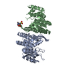
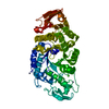
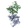
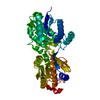
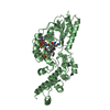
| Title | Structure of epoxyqueuosine reductase from Bacillus subtilis with the Asp134 catalytic loop swung out of the active site. | |||||||||
 Components Components |
| |||||||||
 Keywords Keywords | OXIDOREDUCTASE/RNA / B12 /  cobalamin / iron sulfur cluster / tRNA modifying enzyme / OXIDOREDUCTASE-RNA complex cobalamin / iron sulfur cluster / tRNA modifying enzyme / OXIDOREDUCTASE-RNA complex | |||||||||
| Function / homology |  Function and homology information Function and homology informationepoxyqueuosine reductase / epoxyqueuosine reductase activity / queuosine biosynthetic process / tRNA processing / 4 iron, 4 sulfur cluster binding / metal ion binding / cytoplasmSimilarity search - Function | |||||||||
| Biological species |  Bacillus subtilis (bacteria)Escherichia coli (E. coli) Bacillus subtilis (bacteria)Escherichia coli (E. coli) | |||||||||
| Method | X-RAY DIFFRACTION / SYNCHROTRON / MOLECULAR REPLACEMENT / molecular replacement / Resolution: 2.653 Å | |||||||||
 Authors Authors | Dowling, D.P. / Miles, Z.D. / Kohrer, C. / Maiocco, S.J. / Elliott, S.J. / Bandarian, V. / Drennan, C.L. | |||||||||
| Funding support |  United States, 2items United States, 2items
| |||||||||
 Citation Citation | Journal: Nucleic Acids Res. / Year: 2016 Title: Molecular basis of cobalamin-dependent RNA modification. Authors: Dowling, D.P. / Miles, Z.D. / Kohrer, C. / Maiocco, S.J. / Elliott, S.J. / Bandarian, V. / Drennan, C.L. | |||||||||
| History |
|
- Structure visualization
| Structure viewer | Molecule: MolmilJmol/JSmol |
|---|
- Downloads & links
-Download
| PDBx/mmCIF format | 5t8y.cif.gz | 173.7 KB | Display | PDBx/mmCIF format |
|---|---|---|---|---|
| PDB format | pdb5t8y.ent.gz | 130.6 KB | Display | PDB format |
| PDBx/mmJSON format | 5t8y.json.gz | Tree view | PDBx/mmJSON format | |
| Others |  Other downloads Other downloads |
-Validation report
| Arichive directory | https://data.pdbj.org/pub/pdb/validation_reports/t8/5t8yftp://data.pdbj.org/pub/pdb/validation_reports/t8/5t8y | HTTPS FTP |
|---|
-Related structure data
| Related structure data |  5d08SC  5d0aC  5d0bC S: Starting model for refinement C: citing same article ( |
|---|---|
| Similar structure data |


-Links
 PDBj
PDBj


- Assembly
| Deposited unit | 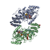
| ||||||||
|---|---|---|---|---|---|---|---|---|---|
| 1 | 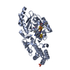
| ||||||||
| 2 | 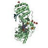
| ||||||||
| Unit cell |
|
-Components
-Protein / RNA chain , 2 types, 3 molecules ABX
| #1: Protein | Mass: 48736.641 Da / Num. of mol.: 2 Source method: isolated from a genetically manipulated source Source: (gene. exp.) Bacillus subtilis (strain 168) (bacteria)Strain: 168 / Gene: queG, ygaP, yhbA, BSU08910 / Production host: Escherichia coli (E. coli) / References: UniProt: P97030, epoxyqueuosine reductase#2: RNA chain | | Mass: 5427.285 Da / Num. of mol.: 1 / Source method: obtained synthetically / Details: anticodon stem loop of Tyr-tRNA from E. coli / Source: (synth.) Escherichia coli (E. coli) |
|---|
-Non-polymers , 4 types, 27 molecules 


| #3: Chemical | ChemComp-SF4 / Iron–sulfur cluster Mass: 351.640 Da / Num. of mol.: 4 / Source method: obtained synthetically / Formula: Fe4S4 Mass: 351.640 Da / Num. of mol.: 4 / Source method: obtained synthetically / Formula: Fe4S4#4: Chemical | Vitamin B12 Mass: 1330.356 Da / Num. of mol.: 2 / Source method: obtained synthetically / Formula: C62H89CoN13O14P Mass: 1330.356 Da / Num. of mol.: 2 / Source method: obtained synthetically / Formula: C62H89CoN13O14P#5: Chemical | ChemComp-PO4 / Phosphate Mass: 94.971 Da / Num. of mol.: 7 / Source method: obtained synthetically / Formula: PO4 Mass: 94.971 Da / Num. of mol.: 7 / Source method: obtained synthetically / Formula: PO4#6: Water | ChemComp-HOH / | WaterMass: 18.015 Da / Num. of mol.: 14 / Source method: isolated from a natural source / Formula: H2O |
|---|
-Experimental details
-Experiment
| Experiment | Method: X-RAY DIFFRACTION / Number of used crystals: 1 |
|---|
- Sample preparation
| Crystal | Density Matthews: 2.29 Å3/Da / Density % sol: 46.25 % |
|---|---|
| Crystal grow | Temperature: 294 K / Method: vapor diffusion, sitting drop / pH: 4.5 Details: 0.1 M acetate (pH 4.5), 0.4 M (NH4)H2PO4, 12% (w/v) PEG 3350 |
-Data collection
| Diffraction | Mean temperature: 100 K |
|---|---|
| Diffraction source | Source: SYNCHROTRON / Site: APS / Beamline: 24-ID-C / Wavelength: 0.9795 Å |
| Detector | Type: DECTRIS PILATUS 6M-F / Detector: PIXEL / Date: Nov 22, 2013 |
| Radiation | Protocol: SINGLE WAVELENGTH / Monochromatic (M) / Laue (L): M / Scattering type: x-ray |
| Radiation wavelength | Wavelength: 0.9795 Å / Relative weight: 1 |
| Reflection | Resolution: 2.65→50 Å / Num. obs: 25487 / % possible obs: 95.1 % / Redundancy: 4.1 % / Rmerge(I) obs: 0.113 / Net I/σ(I): 12 |
| Reflection shell | Resolution: 2.65→2.74 Å / Redundancy: 3.7 % / Rmerge(I) obs: 0.451 / Mean I/σ(I) obs: 1.95 / % possible all: 79 |
-Phasing
| Phasing | Method: molecular replacement |
|---|
- Processing
| Software |
| ||||||||||||||||||||||||||||||||||||||||||||||||||||||||||||||||||||||
|---|---|---|---|---|---|---|---|---|---|---|---|---|---|---|---|---|---|---|---|---|---|---|---|---|---|---|---|---|---|---|---|---|---|---|---|---|---|---|---|---|---|---|---|---|---|---|---|---|---|---|---|---|---|---|---|---|---|---|---|---|---|---|---|---|---|---|---|---|---|---|---|
| Refinement | Method to determine structure: MOLECULAR REPLACEMENT Starting model: 5D08 Resolution: 2.653→47.9 Å / SU ML: 0.41 / Cross valid method: FREE R-VALUE / σ(F): 1.25 / Phase error: 37.27
| ||||||||||||||||||||||||||||||||||||||||||||||||||||||||||||||||||||||
| Solvent computation | Shrinkage radii: 0.9 Å / VDW probe radii: 1.11 Å | ||||||||||||||||||||||||||||||||||||||||||||||||||||||||||||||||||||||
| Displacement parameters | Biso max: 109.46 Å2 / Biso mean: 71.4087 Å2 / Biso min: 58.02 Å2 | ||||||||||||||||||||||||||||||||||||||||||||||||||||||||||||||||||||||
| Refinement step | Cycle: final / Resolution: 2.653→47.9 Å
| ||||||||||||||||||||||||||||||||||||||||||||||||||||||||||||||||||||||
| Refine LS restraints |
| ||||||||||||||||||||||||||||||||||||||||||||||||||||||||||||||||||||||
| LS refinement shell | Refine-ID: X-RAY DIFFRACTION / Total num. of bins used: 9
|
