 Movie
Movie Controller


+ Open data

- Basic information
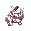
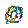
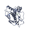
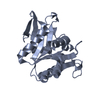
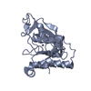
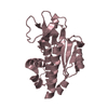
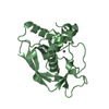
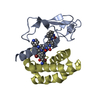
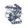
| Entry | Database: PDB / ID: 3wpq | ||||||
|---|---|---|---|---|---|---|---|
| Title | crystal structure of the GAP domain of MgcRacGAP(S387A) | ||||||
 Components Components | Rac GTPase-activating protein 1 | ||||||
 Keywords Keywords |  SIGNALING PROTEIN / GTPase activation / small G-proteins SIGNALING PROTEIN / GTPase activation / small G-proteins | ||||||
| Function / homology |  Function and homology informationcentralspindlin complex / sulfate transport / actomyosin contractile ring assembly / mitotic spindle midzone assembly / regulation of attachment of spindle microtubules to kinetochore / gamma-tubulin binding / Kinesins / RHOD GTPase cycle / Flemming body / regulation of small GTPase mediated signal transduction ...centralspindlin complex / sulfate transport / actomyosin contractile ring assembly / mitotic spindle midzone assembly / regulation of attachment of spindle microtubules to kinetochore / gamma-tubulin binding / Kinesins / RHOD GTPase cycle / Flemming body / regulation of small GTPase mediated signal transduction / COPI-dependent Golgi-to-ER retrograde traffic / RHOB GTPase cycle / beta-tubulin binding / RHOC GTPase cycle / positive regulation of cytokinesis / phosphatidylinositol-3,4,5-trisphosphate binding / cleavage furrow / regulation of embryonic development / mitotic cytokinesis / CDC42 GTPase cycle / Rho protein signal transduction / alpha-tubulin binding / neuroblast proliferation / RHOA GTPase cycle / RAC2 GTPase cycle / RAC3 GTPase cycle / spindle midzone / monoatomic ion transport / RAC1 GTPase cycle / MHC class II antigen presentation / GTPase activator activity / erythrocyte differentiation / acrosomal vesicle / cytoplasmic side of plasma membrane / mitotic spindle / spindle / midbody / microtubule binding / spermatogenesis / microtubule / protein kinase binding / extracellular exosome / nucleoplasm / metal ion binding / nucleus / cytosol Function and homology informationcentralspindlin complex / sulfate transport / actomyosin contractile ring assembly / mitotic spindle midzone assembly / regulation of attachment of spindle microtubules to kinetochore / gamma-tubulin binding / Kinesins / RHOD GTPase cycle / Flemming body / regulation of small GTPase mediated signal transduction ...centralspindlin complex / sulfate transport / actomyosin contractile ring assembly / mitotic spindle midzone assembly / regulation of attachment of spindle microtubules to kinetochore / gamma-tubulin binding / Kinesins / RHOD GTPase cycle / Flemming body / regulation of small GTPase mediated signal transduction / COPI-dependent Golgi-to-ER retrograde traffic / RHOB GTPase cycle / beta-tubulin binding / RHOC GTPase cycle / positive regulation of cytokinesis / phosphatidylinositol-3,4,5-trisphosphate binding / cleavage furrow / regulation of embryonic development / mitotic cytokinesis / CDC42 GTPase cycle / Rho protein signal transduction / alpha-tubulin binding / neuroblast proliferation / RHOA GTPase cycle / RAC2 GTPase cycle / RAC3 GTPase cycle / spindle midzone / monoatomic ion transport / RAC1 GTPase cycle / MHC class II antigen presentation / GTPase activator activity / erythrocyte differentiation / acrosomal vesicle / cytoplasmic side of plasma membrane / mitotic spindle / spindle / midbody / microtubule binding / spermatogenesis / microtubule / protein kinase binding / extracellular exosome / nucleoplasm / metal ion binding / nucleus / cytosolSimilarity search - Function | ||||||
| Biological species |  Homo sapiens (human) Homo sapiens (human) | ||||||
| Method | X-RAY DIFFRACTION / MOLECULAR REPLACEMENT / Resolution: 1.84 Å | ||||||
 Authors Authors | Murayama, K. / Kato-Murayama, M. / Shirouzu, M. / Kitamura, T. / Yokoyama, S. | ||||||
 Citation Citation | Journal: To be Published Title: crystal structure of the GAP domain of MgcRacGAP Authors: Murayama, K. / Kato-Murayama, M. / Shirouzu, M. / Kitamura, T. / Yokoyama, S. | ||||||
| History |
|
- Structure visualization
| Structure viewer | Molecule: MolmilJmol/JSmol |
|---|
- Downloads & links
-Download
| PDBx/mmCIF format | 3wpq.cif.gz | 102.1 KB | Display | PDBx/mmCIF format |
|---|---|---|---|---|
| PDB format | pdb3wpq.ent.gz | 77.8 KB | Display | PDB format |
| PDBx/mmJSON format | 3wpq.json.gz | Tree view | PDBx/mmJSON format | |
| Others |  Other downloads Other downloads |
-Validation report
| Arichive directory | https://data.pdbj.org/pub/pdb/validation_reports/wp/3wpqftp://data.pdbj.org/pub/pdb/validation_reports/wp/3wpq | HTTPS FTP |
|---|
-Related structure data
| Related structure data | 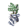 3wpsC  2ovjS C: citing same article ( S: Starting model for refinement |
|---|---|
| Similar structure data |

-Links
 PDBj
PDBj


- Assembly
| Deposited unit | 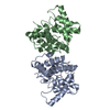
| ||||||||
|---|---|---|---|---|---|---|---|---|---|
| 1 | 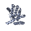
| ||||||||
| 2 | 
| ||||||||
| Unit cell |
|
-Components
| #1: Protein | Mass: 23176.887 Da / Num. of mol.: 2 / Fragment: UNP residues 346-546 / Mutation: S387A Source method: isolated from a genetically manipulated source Source: (gene. exp.) Homo sapiens (human) / Description: PLASMID pCR2.1 / Gene: RACGAP1, KIAA1478, MGCRACGAP / Production host: cell-free protein synthesis (unknown) / References: UniProt: Q9H0H5#2: Water | ChemComp-HOH / | Water Mass: 18.015 Da / Num. of mol.: 551 / Source method: isolated from a natural source / Formula: H2O Mass: 18.015 Da / Num. of mol.: 551 / Source method: isolated from a natural source / Formula: H2OSequence details | THIS SEQUENCE CORRESPOND | |
|---|
-Experimental details
-Experiment
| Experiment | Method: X-RAY DIFFRACTION / Number of used crystals: 1 |
|---|
- Sample preparation
| Crystal | Density Matthews: 2.43 Å3/Da / Density % sol: 49.33 % |
|---|---|
| Crystal grow | Temperature: 293 K / Method: vapor diffusion, sitting drop / pH: 7.5 Details: 1.0M Na Citrate, 0.1M Hepes, pH 7.5, VAPOR DIFFUSION, SITTING DROP, temperature 293K |
-Data collection
| Diffraction | Mean temperature: 100 K |
|---|---|
| Diffraction source | Source: ROTATING ANODE / Type: RIGAKU FR-E SUPERBRIGHT / Wavelength: 1.5418 Å |
| Detector | Type: RIGAKU RAXIS IV++ / Detector: IMAGE PLATE / Date: Apr 24, 2013 |
| Radiation | Protocol: SINGLE WAVELENGTH / Monochromatic (M) / Laue (L): M / Scattering type: x-ray |
| Radiation wavelength | Wavelength: 1.5418 Å / Relative weight: 1 |
| Reflection | Resolution: 1.84→50 Å / Num. obs: 38896 / % possible obs: 100 % / Observed criterion σ(I): -3 / Redundancy: 3.7 % / Biso Wilson estimate: 19.8 Å2 / Rsym value: 0.075 / Net I/σ(I): 17.1 |
| Reflection shell | Resolution: 1.84→1.91 Å / Mean I/σ(I) obs: 3.1 / Rsym value: 0.432 / % possible all: 100 |
- Processing
| Software |
| ||||||||||||||||||||||||||||||||||||||||||||||||||||||||||||||||||||||||||||||||
|---|---|---|---|---|---|---|---|---|---|---|---|---|---|---|---|---|---|---|---|---|---|---|---|---|---|---|---|---|---|---|---|---|---|---|---|---|---|---|---|---|---|---|---|---|---|---|---|---|---|---|---|---|---|---|---|---|---|---|---|---|---|---|---|---|---|---|---|---|---|---|---|---|---|---|---|---|---|---|---|---|---|
| Refinement | Method to determine structure: MOLECULAR REPLACEMENT Starting model: 2OVJ Resolution: 1.84→28.89 Å / Rfactor Rfree error: 0.005 / Data cutoff high absF: 1027008.88 / Data cutoff low absF: 0 / Isotropic thermal model: RESTRAINED / Cross valid method: THROUGHOUT / σ(F): 0 / Stereochemistry target values: Engh & Huber
| ||||||||||||||||||||||||||||||||||||||||||||||||||||||||||||||||||||||||||||||||
| Solvent computation | Solvent model: FLAT MODEL / Bsol: 57.8917 Å2 / ksol: 0.390357 e/Å3 | ||||||||||||||||||||||||||||||||||||||||||||||||||||||||||||||||||||||||||||||||
| Displacement parameters | Biso mean: 22.5 Å2
| ||||||||||||||||||||||||||||||||||||||||||||||||||||||||||||||||||||||||||||||||
| Refine analyze |
| ||||||||||||||||||||||||||||||||||||||||||||||||||||||||||||||||||||||||||||||||
| Refinement step | Cycle: LAST / Resolution: 1.84→28.89 Å
| ||||||||||||||||||||||||||||||||||||||||||||||||||||||||||||||||||||||||||||||||
| Refine LS restraints |
| ||||||||||||||||||||||||||||||||||||||||||||||||||||||||||||||||||||||||||||||||
| LS refinement shell | Resolution: 1.84→1.96 Å / Rfactor Rfree error: 0.015 / Total num. of bins used: 6
| ||||||||||||||||||||||||||||||||||||||||||||||||||||||||||||||||||||||||||||||||
| Xplor file |
|
