 Movie
Movie Controller

[English] 日本語
 Yorodumi
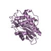
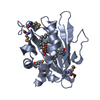
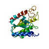
- PDB-2ovj: The crystal structure of the human Rac GTPase activating protein ... -
+ Open data

- Basic information
| Entry | Database: PDB / ID: 2ovj | ||||||
|---|---|---|---|---|---|---|---|
| Title | The crystal structure of the human Rac GTPase activating protein 1 (RACGAP1) MgcRacGAP. | ||||||
 Components Components | Rac GTPase-activating protein 1 | ||||||
 Keywords Keywords |  SIGNALING PROTEIN / Structural Genomics / Structural Genomics Consortium / SGC SIGNALING PROTEIN / Structural Genomics / Structural Genomics Consortium / SGC | ||||||
| Function / homology |  Function and homology informationcentralspindlin complex / actomyosin contractile ring assembly / sulfate transport / mitotic spindle midzone assembly / regulation of attachment of spindle microtubules to kinetochore / gamma-tubulin binding / RHOD GTPase cycle / Kinesins / Flemming body / regulation of small GTPase mediated signal transduction ...centralspindlin complex / actomyosin contractile ring assembly / sulfate transport / mitotic spindle midzone assembly / regulation of attachment of spindle microtubules to kinetochore / gamma-tubulin binding / RHOD GTPase cycle / Kinesins / Flemming body / regulation of small GTPase mediated signal transduction / COPI-dependent Golgi-to-ER retrograde traffic / RHOB GTPase cycle / beta-tubulin binding / RHOC GTPase cycle / positive regulation of cytokinesis / phosphatidylinositol-3,4,5-trisphosphate binding / cleavage furrow / regulation of embryonic development / CDC42 GTPase cycle / Rho protein signal transduction / mitotic cytokinesis / alpha-tubulin binding / neuroblast proliferation / RHOA GTPase cycle / RAC2 GTPase cycle / RAC3 GTPase cycle / spindle midzone / monoatomic ion transport / RAC1 GTPase cycle / MHC class II antigen presentation / GTPase activator activity / erythrocyte differentiation / acrosomal vesicle / mitotic spindle / cytoplasmic side of plasma membrane / spindle / protein-macromolecule adaptor activity / midbody / microtubule binding / spermatogenesis / microtubule / protein kinase binding / extracellular exosome / nucleoplasm / metal ion binding / nucleus / cytosol Function and homology informationcentralspindlin complex / actomyosin contractile ring assembly / sulfate transport / mitotic spindle midzone assembly / regulation of attachment of spindle microtubules to kinetochore / gamma-tubulin binding / RHOD GTPase cycle / Kinesins / Flemming body / regulation of small GTPase mediated signal transduction ...centralspindlin complex / actomyosin contractile ring assembly / sulfate transport / mitotic spindle midzone assembly / regulation of attachment of spindle microtubules to kinetochore / gamma-tubulin binding / RHOD GTPase cycle / Kinesins / Flemming body / regulation of small GTPase mediated signal transduction / COPI-dependent Golgi-to-ER retrograde traffic / RHOB GTPase cycle / beta-tubulin binding / RHOC GTPase cycle / positive regulation of cytokinesis / phosphatidylinositol-3,4,5-trisphosphate binding / cleavage furrow / regulation of embryonic development / CDC42 GTPase cycle / Rho protein signal transduction / mitotic cytokinesis / alpha-tubulin binding / neuroblast proliferation / RHOA GTPase cycle / RAC2 GTPase cycle / RAC3 GTPase cycle / spindle midzone / monoatomic ion transport / RAC1 GTPase cycle / MHC class II antigen presentation / GTPase activator activity / erythrocyte differentiation / acrosomal vesicle / mitotic spindle / cytoplasmic side of plasma membrane / spindle / protein-macromolecule adaptor activity / midbody / microtubule binding / spermatogenesis / microtubule / protein kinase binding / extracellular exosome / nucleoplasm / metal ion binding / nucleus / cytosolSimilarity search - Function | ||||||
| Biological species |  Homo sapiens (human) Homo sapiens (human) | ||||||
| Method | X-RAY DIFFRACTION / SYNCHROTRON / MOLECULAR REPLACEMENT / Resolution: 1.49 Å | ||||||
 Authors Authors | Shrestha, L. / Papagrigoriou, E. / Soundararajan, M. / Elkins, J. / Johansson, C. / von Delft, F. / Pike, A.C.W. / Burgess, N. / Turnbull, A. / Debreczeni, J. ...Shrestha, L. / Papagrigoriou, E. / Soundararajan, M. / Elkins, J. / Johansson, C. / von Delft, F. / Pike, A.C.W. / Burgess, N. / Turnbull, A. / Debreczeni, J. / Gorrec, F. / Umeano, C. / Edwards, A. / Arrowsmith, C.H. / Weigelt, J. / Sundstrom, M. / Doyle, D.A. / Structural Genomics Consortium (SGC) | ||||||
 Citation Citation | Journal: To be Published Title: The crystal structure of the human Rac GTPase activating protein 1 (RACGAP1) MgcRacGAP. Authors: Shrestha, L. / Papagrigoriou, E. / Soundararajan, M. / Elkins, J. / Johansson, C. / von Delft, F. / Pike, A.C.W. / Burgess, N. / Turnbull, A. / Debreczeni, J. / Gorrec, F. / Umeano, C. / ...Authors: Shrestha, L. / Papagrigoriou, E. / Soundararajan, M. / Elkins, J. / Johansson, C. / von Delft, F. / Pike, A.C.W. / Burgess, N. / Turnbull, A. / Debreczeni, J. / Gorrec, F. / Umeano, C. / Edwards, A. / Arrowsmith, C.H. / Weigelt, J. / Sundstrom, M. / Doyle, D.A. | ||||||
| History |
|
- Structure visualization
| Structure viewer | Molecule: MolmilJmol/JSmol |
|---|
- Downloads & links
-Download
| PDBx/mmCIF format | 2ovj.cif.gz | 59 KB | Display | PDBx/mmCIF format |
|---|---|---|---|---|
| PDB format | pdb2ovj.ent.gz | 41.3 KB | Display | PDB format |
| PDBx/mmJSON format | 2ovj.json.gz | Tree view | PDBx/mmJSON format | |
| Others |  Other downloads Other downloads |
-Validation report
| Arichive directory | https://data.pdbj.org/pub/pdb/validation_reports/ov/2ovjftp://data.pdbj.org/pub/pdb/validation_reports/ov/2ovj | HTTPS FTP |
|---|
-Related structure data
| Related structure data |  1f7cS S: Starting model for refinement |
|---|---|
| Similar structure data |


-Links
 PDBj
PDBj


- Assembly
| Deposited unit | 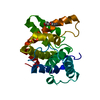
| ||||||||
|---|---|---|---|---|---|---|---|---|---|
| 1 |
| ||||||||
| Unit cell |
| ||||||||
| Details | The biological unit is a monomer |
-Components
| #1: Protein | Mass: 22747.562 Da / Num. of mol.: 1 / Fragment: Rho-GAP domain Source method: isolated from a genetically manipulated source Source: (gene. exp.) Homo sapiens (human) / Gene: RACGAP1, KIAA1478, MGCRACGAP / Plasmid: pNIC28-Bsa4 / Species (production host): Escherichia coli / Production host:  Escherichia coli BL21(DE3) (bacteria) / Strain (production host): BL21(DE3) / References: UniProt: Q9H0H5 Escherichia coli BL21(DE3) (bacteria) / Strain (production host): BL21(DE3) / References: UniProt: Q9H0H5 |
|---|---|
| #2: Chemical | ChemComp-7PE / Polyethylene glycol  Mass: 310.384 Da / Num. of mol.: 1 / Source method: obtained synthetically / Formula: C14H30O7 Mass: 310.384 Da / Num. of mol.: 1 / Source method: obtained synthetically / Formula: C14H30O7 |
| #3: Water | ChemComp-HOH / Water Mass: 18.015 Da / Num. of mol.: 171 / Source method: isolated from a natural source / Formula: H2O Mass: 18.015 Da / Num. of mol.: 171 / Source method: isolated from a natural source / Formula: H2O |
-Experimental details
-Experiment
| Experiment | Method: X-RAY DIFFRACTION / Number of used crystals: 1 |
|---|
- Sample preparation
| Crystal | Density Matthews: 2.12 Å3/Da / Density % sol: 41.86 % |
|---|---|
| Crystal grow | Temperature: 298 K / Method: vapor diffusion, sitting drop / pH: 5.5 Details: 25% PEG 3350, 0.1M BIS-TRIS, pH 5.5, VAPOR DIFFUSION, SITTING DROP, temperature 298K |
-Data collection
| Diffraction | Mean temperature: 100 K |
|---|---|
| Diffraction source | Source: SYNCHROTRON / Site: SLS  / Beamline: X10SA / Wavelength: 1 Å / Beamline: X10SA / Wavelength: 1 Å |
| Detector | Type: MARMOSAIC 225 mm CCD / Detector: CCD / Date: Dec 16, 2006 |
| Radiation | Monochromator: Si(111) / Protocol: SINGLE WAVELENGTH / Monochromatic (M) / Laue (L): M / Scattering type: x-ray |
| Radiation wavelength | Wavelength: 1 Å / Relative weight: 1 |
| Reflection | Resolution: 1.49→42.07 Å / Num. all: 32274 / Num. obs: 32274 / % possible obs: 99.9 % / Observed criterion σ(F): 0 / Observed criterion σ(I): 0 / Redundancy: 7 % / Rmerge(I) obs: 0.075 / Rsym value: 0.075 |
| Reflection shell | Resolution: 1.49→1.54 Å / Redundancy: 5.5 % / Rmerge(I) obs: 0.491 / Num. unique all: 3134 / % possible all: 99.3 |
- Processing
| Software |
| ||||||||||||||||||||||||||||||||||||||||||||||||||||||||||||||||||||||||||||||||||||||||||||||||||||||||||||||||||||||||||||||||||||||||||||||||||||||||||||||||||||||||||
|---|---|---|---|---|---|---|---|---|---|---|---|---|---|---|---|---|---|---|---|---|---|---|---|---|---|---|---|---|---|---|---|---|---|---|---|---|---|---|---|---|---|---|---|---|---|---|---|---|---|---|---|---|---|---|---|---|---|---|---|---|---|---|---|---|---|---|---|---|---|---|---|---|---|---|---|---|---|---|---|---|---|---|---|---|---|---|---|---|---|---|---|---|---|---|---|---|---|---|---|---|---|---|---|---|---|---|---|---|---|---|---|---|---|---|---|---|---|---|---|---|---|---|---|---|---|---|---|---|---|---|---|---|---|---|---|---|---|---|---|---|---|---|---|---|---|---|---|---|---|---|---|---|---|---|---|---|---|---|---|---|---|---|---|---|---|---|---|---|---|---|---|
| Refinement | Method to determine structure: MOLECULAR REPLACEMENT Starting model: PDB entry 1F7C Resolution: 1.49→42.07 Å / Cor.coef. Fo:Fc: 0.968 / Cor.coef. Fo:Fc free: 0.96 / SU B: 1.956 / SU ML: 0.037 / TLS residual ADP flag: LIKELY RESIDUAL / Cross valid method: THROUGHOUT / σ(F): 0 / σ(I): 0 / ESU R: 0.064 / ESU R Free: 0.065 / Stereochemistry target values: MAXIMUM LIKELIHOOD / Details: HYDROGENS HAVE BEEN ADDED IN THE RIDING POSITIONS
| ||||||||||||||||||||||||||||||||||||||||||||||||||||||||||||||||||||||||||||||||||||||||||||||||||||||||||||||||||||||||||||||||||||||||||||||||||||||||||||||||||||||||||
| Solvent computation | Ion probe radii: 0.8 Å / Shrinkage radii: 0.8 Å / VDW probe radii: 1.4 Å / Solvent model: BABINET MODEL WITH MASK | ||||||||||||||||||||||||||||||||||||||||||||||||||||||||||||||||||||||||||||||||||||||||||||||||||||||||||||||||||||||||||||||||||||||||||||||||||||||||||||||||||||||||||
| Displacement parameters | Biso mean: 11.767 Å2
| ||||||||||||||||||||||||||||||||||||||||||||||||||||||||||||||||||||||||||||||||||||||||||||||||||||||||||||||||||||||||||||||||||||||||||||||||||||||||||||||||||||||||||
| Refinement step | Cycle: LAST / Resolution: 1.49→42.07 Å
| ||||||||||||||||||||||||||||||||||||||||||||||||||||||||||||||||||||||||||||||||||||||||||||||||||||||||||||||||||||||||||||||||||||||||||||||||||||||||||||||||||||||||||
| Refine LS restraints |
| ||||||||||||||||||||||||||||||||||||||||||||||||||||||||||||||||||||||||||||||||||||||||||||||||||||||||||||||||||||||||||||||||||||||||||||||||||||||||||||||||||||||||||
| LS refinement shell | Resolution: 1.49→1.53 Å / Total num. of bins used: 20
| ||||||||||||||||||||||||||||||||||||||||||||||||||||||||||||||||||||||||||||||||||||||||||||||||||||||||||||||||||||||||||||||||||||||||||||||||||||||||||||||||||||||||||
| Refinement TLS params. | Method: refined / Origin x: 13.3898 Å / Origin y: 1.8942 Å / Origin z: 12.3309 Å
|
