 Movie
Movie Controller


+ Open data

- Basic information
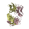
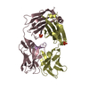
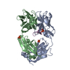
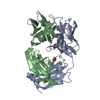
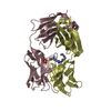
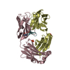
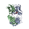
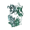
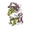
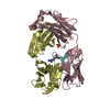
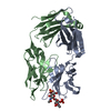
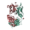
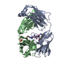
| Entry | Database: PDB / ID: 5t1k | |||||||||
|---|---|---|---|---|---|---|---|---|---|---|
| Title | Cetuximab Fab in complex with CQFDA(Ph)2STRRLKC | |||||||||
 Components Components |
| |||||||||
 Keywords Keywords |  IMMUNE SYSTEM / antibody / anti-EGFR IMMUNE SYSTEM / antibody / anti-EGFR | |||||||||
| Function / homology | Immunoglobulins / Immunoglobulin-like / Sandwich / Mainly Beta / MESO-ERYTHRITOL / PHOSPHATE ION Function and homology information Function and homology information | |||||||||
| Biological species |  Mus musculus (house mouse) Mus musculus (house mouse) Homo sapiens (human) Homo sapiens (human)synthetic construct (others) | |||||||||
| Method | X-RAY DIFFRACTION / MOLECULAR REPLACEMENT / Resolution: 2.48 Å | |||||||||
 Authors Authors | Bzymek, K.P. / Williams, J.C. | |||||||||
 Citation Citation | Journal: Acta Crystallogr F Struct Biol Commun / Year: 2016 Title: Natural and non-natural amino-acid side-chain substitutions: affinity and diffraction studies of meditope-Fab complexes. Authors: Bzymek, K.P. / Avery, K.A. / Ma, Y. / Horne, D.A. / Williams, J.C. | |||||||||
| History |
|
- Structure visualization
| Structure viewer | Molecule: MolmilJmol/JSmol |
|---|
- Downloads & links
-Download
| PDBx/mmCIF format | 5t1k.cif.gz | 196.2 KB | Display | PDBx/mmCIF format |
|---|---|---|---|---|
| PDB format | pdb5t1k.ent.gz | 153.9 KB | Display | PDB format |
| PDBx/mmJSON format | 5t1k.json.gz | Tree view | PDBx/mmJSON format | |
| Others |  Other downloads Other downloads |
-Validation report
| Arichive directory | https://data.pdbj.org/pub/pdb/validation_reports/t1/5t1kftp://data.pdbj.org/pub/pdb/validation_reports/t1/5t1k | HTTPS FTP |
|---|
-Related structure data
| Related structure data | 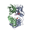 5etuC 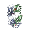 5eukC 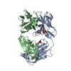 5f88C  5ff6C 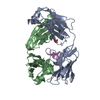 5i2iC 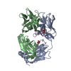 5iopC 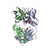 5ir1C 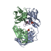 5itfC 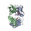 5iv2C 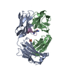 5ivzC 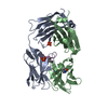 5t1lC 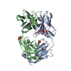 5t1mC 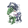 5th2C  4gw1S  4iwe S: Starting model for refinement C: citing same article ( |
|---|---|
| Similar structure data |


-Links
 PDBj
PDBj

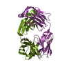
- Assembly
| Deposited unit | 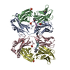
| ||||||||
|---|---|---|---|---|---|---|---|---|---|
| 1 | 
| ||||||||
| 2 | 
| ||||||||
| Unit cell |
|
-Components
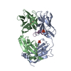
-Protein/peptide , 1 types, 2 molecules EF
| #4: Protein/peptide | Mass: 1582.868 Da / Num. of mol.: 2 / Source method: obtained synthetically / Source: (synth.) synthetic construct (others) |
|---|
-Antibody , 3 types, 4 molecules ACBD
| #1: Antibody | Mass: 23287.705 Da / Num. of mol.: 2 Source method: isolated from a genetically manipulated source Source: (gene. exp.) Mus musculus, Homo sapiens / Production host: Mus musculus (house mouse)#2: Antibody | | Mass: 23708.475 Da / Num. of mol.: 1 Source method: isolated from a genetically manipulated source Source: (gene. exp.) Mus musculus, Homo sapiens / Production host: Mus musculus (house mouse)#3: Antibody | | Mass: 23725.504 Da / Num. of mol.: 1 Source method: isolated from a genetically manipulated source Source: (gene. exp.) Mus musculus, Homo sapiens / Production host: Mus musculus (house mouse) |
|---|
-Non-polymers , 3 types, 524 molecules 
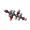

| #5: Chemical | ChemComp-PO4 / Phosphate Mass: 94.971 Da / Num. of mol.: 5 / Source method: obtained synthetically / Formula: PO4 Mass: 94.971 Da / Num. of mol.: 5 / Source method: obtained synthetically / Formula: PO4#6: Chemical | ChemComp-MRY / | Erythritol Mass: 122.120 Da / Num. of mol.: 1 / Source method: obtained synthetically / Formula: C4H10O4 Mass: 122.120 Da / Num. of mol.: 1 / Source method: obtained synthetically / Formula: C4H10O4#7: Water | ChemComp-HOH / | WaterMass: 18.015 Da / Num. of mol.: 518 / Source method: isolated from a natural source / Formula: H2O |
|---|
-Experimental details
-Experiment
| Experiment | Method: X-RAY DIFFRACTION / Number of used crystals: 1 |
|---|
- Sample preparation
| Crystal | Density Matthews: 2.92 Å3/Da / Density % sol: 57.92 % |
|---|---|
| Crystal grow | Temperature: 293 K / Method: vapor diffusion, hanging drop / pH: 5.5 Details: 0.1 CITRIC ACID, 0.1 M SODIUM HYDROGEN PHOSPHATE, 0.5 M POTASSIUM HYDROGEN PHOSPHATE, 1.6 M SODIUM DIHYDROGEN PHOSPHATE, PH 5.5 PH range: 5.5 |
-Data collection
| Diffraction | Mean temperature: 100 K |
|---|---|
| Diffraction source | Source: ROTATING ANODE / Type: RIGAKU MICROMAX-007 HF / Wavelength: 1.5418 Å |
| Detector | Type: RIGAKU RAXIS IV++ / Detector: IMAGE PLATE / Date: Sep 2, 2011 |
| Radiation | Monochromator: VARIMAX / Protocol: SINGLE WAVELENGTH / Monochromatic (M) / Laue (L): M / Scattering type: x-ray |
| Radiation wavelength | Wavelength: 1.5418 Å / Relative weight: 1 |
| Reflection | Resolution: 2.48→33.2 Å / Num. obs: 41117 / % possible obs: 99.1 % / Observed criterion σ(I): 2 / Redundancy: 4.6 % / Rmerge(I) obs: 0.053 / Net I/σ(I): 23.5 |
| Reflection shell | Resolution: 2.48→2.54 Å / Redundancy: 3.2 % / Rmerge(I) obs: 0.248 / Mean I/σ(I) obs: 4.7 / % possible all: 90.3 |
- Processing
| Software |
| ||||||||||||||||||||||||||||||||||||||||||||||||||||||||||||||||||||||||||||||||||||||||||||||||||||||||||||||||
|---|---|---|---|---|---|---|---|---|---|---|---|---|---|---|---|---|---|---|---|---|---|---|---|---|---|---|---|---|---|---|---|---|---|---|---|---|---|---|---|---|---|---|---|---|---|---|---|---|---|---|---|---|---|---|---|---|---|---|---|---|---|---|---|---|---|---|---|---|---|---|---|---|---|---|---|---|---|---|---|---|---|---|---|---|---|---|---|---|---|---|---|---|---|---|---|---|---|---|---|---|---|---|---|---|---|---|---|---|---|---|---|---|---|
| Refinement | Method to determine structure: MOLECULAR REPLACEMENT Starting model: 4gw1 Resolution: 2.48→33.2 Å / SU ML: 0.25 / Cross valid method: FREE R-VALUE / σ(F): 2 / Phase error: 20.11
| ||||||||||||||||||||||||||||||||||||||||||||||||||||||||||||||||||||||||||||||||||||||||||||||||||||||||||||||||
| Solvent computation | Shrinkage radii: 0.9 Å / VDW probe radii: 1.2 Å | ||||||||||||||||||||||||||||||||||||||||||||||||||||||||||||||||||||||||||||||||||||||||||||||||||||||||||||||||
| Refinement step | Cycle: LAST / Resolution: 2.48→33.2 Å
| ||||||||||||||||||||||||||||||||||||||||||||||||||||||||||||||||||||||||||||||||||||||||||||||||||||||||||||||||
| Refine LS restraints |
| ||||||||||||||||||||||||||||||||||||||||||||||||||||||||||||||||||||||||||||||||||||||||||||||||||||||||||||||||
| LS refinement shell |
|
