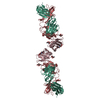
 Movie
Movie Controller

[English] 日本語
 Yorodumi
- PDB-5szp: Protocadherin Gamma B7 extracellular cadherin domains 1-4 P21 cry... -
+ Open data

- Basic information
| Entry | Database: PDB / ID: 5szp | ||||||||||||
|---|---|---|---|---|---|---|---|---|---|---|---|---|---|
| Title | Protocadherin Gamma B7 extracellular cadherin domains 1-4 P21 crystal form | ||||||||||||
 Components Components | Protocadherin Gamma B7 | ||||||||||||
 Keywords Keywords |  CELL ADHESION CELL ADHESION | ||||||||||||
| Function / homology |  Function and homology information Function and homology informationhomophilic cell adhesion via plasma membrane adhesion molecules / cell adhesion / calcium ion binding / membrane / plasma membraneSimilarity search - Function | ||||||||||||
| Biological species |  Mus musculus (house mouse) Mus musculus (house mouse) | ||||||||||||
| Method | X-RAY DIFFRACTION / SYNCHROTRON / MOLECULAR REPLACEMENT / Resolution: 3.1 Å | ||||||||||||
 Authors Authors | Goodman, K.M. / Mannepalli, S. / Bahna, F. / Honig, B. / Shapiro, L. | ||||||||||||
| Funding support |  United States, 3items United States, 3items
| ||||||||||||
 Citation Citation | Journal: Elife / Year: 2016 Title: gamma-Protocadherin structural diversity and functional implications. Authors: Goodman, K.M. / Rubinstein, R. / Thu, C.A. / Mannepalli, S. / Bahna, F. / Ahlsen, G. / Rittenhouse, C. / Maniatis, T. / Honig, B. / Shapiro, L. | ||||||||||||
| History |
|
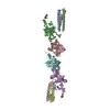
- Structure visualization
| Structure viewer | Molecule: MolmilJmol/JSmol |
|---|
- Downloads & links
-Download
| PDBx/mmCIF format | 5szp.cif.gz | 325.3 KB | Display | PDBx/mmCIF format |
|---|---|---|---|---|
| PDB format | pdb5szp.ent.gz | 262.8 KB | Display | PDB format |
| PDBx/mmJSON format | 5szp.json.gz | Tree view | PDBx/mmJSON format | |
| Others |  Other downloads Other downloads |
-Validation report
| Arichive directory | https://data.pdbj.org/pub/pdb/validation_reports/sz/5szpftp://data.pdbj.org/pub/pdb/validation_reports/sz/5szp | HTTPS FTP |
|---|
-Related structure data
| Related structure data |  5szlC  5szmC  5sznC  5szoSC  5szqC  5szrC  5t9tC C: citing same article ( S: Starting model for refinement |
|---|---|
| Similar structure data |

-Links
 PDBj
PDBj


- Assembly
| Deposited unit | 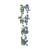
| ||||||||
|---|---|---|---|---|---|---|---|---|---|
| 1 |
| ||||||||
| Unit cell |
|
-Components
-Protein , 1 types, 2 molecules AB
| #1: Protein | Mass: 47295.547 Da / Num. of mol.: 2 Source method: isolated from a genetically manipulated source Source: (gene. exp.) Mus musculus (house mouse) / Gene: Pcdhgb7, mCG_133388 / Cell line (production host): HEK293F / Production host:  Homo sapiens (human) / References: UniProt: Q91XX3 Homo sapiens (human) / References: UniProt: Q91XX3 |
|---|
-Sugars , 2 types, 8 molecules 

| #3: Sugar | ChemComp-MAN / Mannose Type: D-saccharide, alpha linking / Mass: 180.156 Da / Num. of mol.: 7 Type: D-saccharide, alpha linking / Mass: 180.156 Da / Num. of mol.: 7Source method: isolated from a genetically manipulated source Formula: C6H12O6 #4: Sugar | ChemComp-NAG / | N-Acetylglucosamine Type: D-saccharide, beta linking / Mass: 221.208 Da / Num. of mol.: 1 Type: D-saccharide, beta linking / Mass: 221.208 Da / Num. of mol.: 1Source method: isolated from a genetically manipulated source Formula: C8H15NO6 |
|---|
-Non-polymers , 3 types, 24 molecules 

| #2: Chemical | ChemComp-CA /  Mass: 40.078 Da / Num. of mol.: 18 / Source method: obtained synthetically / Formula: Ca Mass: 40.078 Da / Num. of mol.: 18 / Source method: obtained synthetically / Formula: Ca#5: Chemical | ChemComp-TMO / | Trimethylamine N-oxide Mass: 75.110 Da / Num. of mol.: 1 / Source method: obtained synthetically / Formula: C3H9NO Mass: 75.110 Da / Num. of mol.: 1 / Source method: obtained synthetically / Formula: C3H9NO#6: Water | ChemComp-HOH / | WaterMass: 18.015 Da / Num. of mol.: 5 / Source method: isolated from a natural source / Formula: H2O |
|---|
-Experimental details
-Experiment
| Experiment | Method: X-RAY DIFFRACTION / Number of used crystals: 1 |
|---|
- Sample preparation
| Crystal | Density Matthews: 2.55 Å3/Da / Density % sol: 51.68 % / Description: Plate |
|---|---|
| Crystal grow | Temperature: 295 K / Method: batch mode / pH: 8.5 Details: 0.1 M Tris-Cl pH 8.5, 0.2 trimethylamine N-oxide, 5% (v/v) Jeffamine M-600 pH 7.0, 17% (w/v) PEG2000MME |
-Data collection
| Diffraction | Mean temperature: 100 K |
|---|---|
| Diffraction source | Source: SYNCHROTRON / Site: APS / Beamline: 24-ID-C / Wavelength: 0.97919 Å |
| Detector | Type: DECTRIS PILATUS 6M-F / Detector: PIXEL / Date: Jul 22, 2016 |
| Radiation | Protocol: SINGLE WAVELENGTH / Monochromatic (M) / Laue (L): M / Scattering type: x-ray |
| Radiation wavelength | Wavelength: 0.97919 Å / Relative weight: 1 |
| Reflection | Resolution: 3.1→39.95 Å / Num. obs: 17677 / % possible obs: 99.5 % / Redundancy: 3.4 % / Biso Wilson estimate: 108.83 Å2 / CC1/2: 0.982 / Rmerge(I) obs: 0.18 / Net I/σ(I): 5.5 |
| Reflection shell | Resolution: 3.1→3.31 Å / Redundancy: 3.3 % / Rmerge(I) obs: 1.512 / Mean I/σ(I) obs: 0.8 / CC1/2: 0.585 / % possible all: 99.2 |
- Processing
| Software |
| |||||||||||||||||||||||||||||||||||||||||||||||||||||||||||||||||||||||||||||||||||||||||||||||||||||||||||||||||||||||||||||||||||||||||||||||||||||||||||||||||||||||||||||||||||||||||||||||||||||||||||||||||||||||||||||||||
|---|---|---|---|---|---|---|---|---|---|---|---|---|---|---|---|---|---|---|---|---|---|---|---|---|---|---|---|---|---|---|---|---|---|---|---|---|---|---|---|---|---|---|---|---|---|---|---|---|---|---|---|---|---|---|---|---|---|---|---|---|---|---|---|---|---|---|---|---|---|---|---|---|---|---|---|---|---|---|---|---|---|---|---|---|---|---|---|---|---|---|---|---|---|---|---|---|---|---|---|---|---|---|---|---|---|---|---|---|---|---|---|---|---|---|---|---|---|---|---|---|---|---|---|---|---|---|---|---|---|---|---|---|---|---|---|---|---|---|---|---|---|---|---|---|---|---|---|---|---|---|---|---|---|---|---|---|---|---|---|---|---|---|---|---|---|---|---|---|---|---|---|---|---|---|---|---|---|---|---|---|---|---|---|---|---|---|---|---|---|---|---|---|---|---|---|---|---|---|---|---|---|---|---|---|---|---|---|---|---|---|---|---|---|---|---|---|---|---|---|---|---|---|---|---|---|---|
| Refinement | Method to determine structure: MOLECULAR REPLACEMENT Starting model: 5SZO Resolution: 3.1→19.977 Å / SU ML: 0.52 / Cross valid method: FREE R-VALUE / σ(F): 1.33 / Phase error: 36.62
| |||||||||||||||||||||||||||||||||||||||||||||||||||||||||||||||||||||||||||||||||||||||||||||||||||||||||||||||||||||||||||||||||||||||||||||||||||||||||||||||||||||||||||||||||||||||||||||||||||||||||||||||||||||||||||||||||
| Solvent computation | Shrinkage radii: 0.9 Å / VDW probe radii: 1.11 Å | |||||||||||||||||||||||||||||||||||||||||||||||||||||||||||||||||||||||||||||||||||||||||||||||||||||||||||||||||||||||||||||||||||||||||||||||||||||||||||||||||||||||||||||||||||||||||||||||||||||||||||||||||||||||||||||||||
| Refinement step | Cycle: LAST / Resolution: 3.1→19.977 Å
| |||||||||||||||||||||||||||||||||||||||||||||||||||||||||||||||||||||||||||||||||||||||||||||||||||||||||||||||||||||||||||||||||||||||||||||||||||||||||||||||||||||||||||||||||||||||||||||||||||||||||||||||||||||||||||||||||
| Refine LS restraints |
| |||||||||||||||||||||||||||||||||||||||||||||||||||||||||||||||||||||||||||||||||||||||||||||||||||||||||||||||||||||||||||||||||||||||||||||||||||||||||||||||||||||||||||||||||||||||||||||||||||||||||||||||||||||||||||||||||
| LS refinement shell |
| |||||||||||||||||||||||||||||||||||||||||||||||||||||||||||||||||||||||||||||||||||||||||||||||||||||||||||||||||||||||||||||||||||||||||||||||||||||||||||||||||||||||||||||||||||||||||||||||||||||||||||||||||||||||||||||||||
| Refinement TLS params. | Method: refined / Refine-ID: X-RAY DIFFRACTION
| |||||||||||||||||||||||||||||||||||||||||||||||||||||||||||||||||||||||||||||||||||||||||||||||||||||||||||||||||||||||||||||||||||||||||||||||||||||||||||||||||||||||||||||||||||||||||||||||||||||||||||||||||||||||||||||||||
| Refinement TLS group |
|
