 Movie
Movie Controller

[English] 日本語
 Yorodumi
- PDB-5suz: Domain-swapped dimer of human Dishevelled2 DEP domain: C-centered... -
+ Open data

- Basic information
| Entry | Database: PDB / ID: 5suz | ||||||
|---|---|---|---|---|---|---|---|
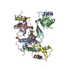
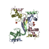
| Title | Domain-swapped dimer of human Dishevelled2 DEP domain: C-centered monoclinic crystal form crystallised from monomeric fraction | ||||||
 Components Components | Segment polarity protein dishevelled homolog DVL-2 | ||||||
 Keywords Keywords |  SIGNALING PROTEIN / Dishevelled / DEP domain / WNT signalling SIGNALING PROTEIN / Dishevelled / DEP domain / WNT signalling | ||||||
| Function / homology |  Function and homology information Function and homology informationNegative regulation of TCF-dependent signaling by DVL-interacting proteins / convergent extension involved in neural plate elongation / planar cell polarity pathway involved in neural tube closure / cochlea morphogenesis / segment specification / WNT5A-dependent internalization of FZD4 / non-canonical Wnt signaling pathway / positive regulation of neuron projection arborization / WNT5:FZD7-mediated leishmania damping / clathrin-coated endocytic vesicle ...Negative regulation of TCF-dependent signaling by DVL-interacting proteins / convergent extension involved in neural plate elongation / planar cell polarity pathway involved in neural tube closure / cochlea morphogenesis / segment specification / WNT5A-dependent internalization of FZD4 / non-canonical Wnt signaling pathway / positive regulation of neuron projection arborization / WNT5:FZD7-mediated leishmania damping / clathrin-coated endocytic vesicle / frizzled binding / PCP/CE pathway / Signaling by Hippo / WNT mediated activation of DVL / aggresome / Disassembly of the destruction complex and recruitment of AXIN to the membrane / Wnt signaling pathway, planar cell polarity pathway / heart looping / outflow tract morphogenesis / lateral plasma membrane / canonical Wnt signaling pathway / positive regulation of JUN kinase activity / Asymmetric localization of PCP proteins / TCF dependent signaling in response to WNT / neural tube closure / RHO GTPases Activate Formins / Degradation of DVL / regulation of actin cytoskeleton organization / positive regulation of JNK cascade / protein localization / small GTPase binding / positive regulation of DNA-binding transcription factor activity / : / protein-macromolecule adaptor activity / Cargo recognition for clathrin-mediated endocytosis / apical part of cell / Clathrin-mediated endocytosis / heart development / regulation of cell population proliferation / cytoplasmic vesicle / nuclear body / intracellular signal transduction / positive regulation of protein phosphorylation / protein domain specific binding / regulation of DNA-templated transcription / protein kinase binding / positive regulation of transcription by RNA polymerase II / nucleoplasm / identical protein binding / nucleus / cytosol / cytoplasmSimilarity search - Function | ||||||
| Biological species |  Homo sapiens (human) Homo sapiens (human) | ||||||
| Method | X-RAY DIFFRACTION / SYNCHROTRON / SAD / Resolution: 1.84 Å | ||||||
 Authors Authors | Renko, M. / Gammons, M.V. / Bienz, M. | ||||||
 Citation Citation | Journal: Mol.Cell / Year: 2016 Title: Wnt Signalosome Assembly by DEP Domain Swapping of Dishevelled. Authors: Gammons, M.V. / Renko, M. / Johnson, C.M. / Rutherford, T.J. / Bienz, M. | ||||||
| History |
|
- Structure visualization
| Structure viewer | Molecule: MolmilJmol/JSmol |
|---|
- Downloads & links
-Download
| PDBx/mmCIF format | 5suz.cif.gz | 86.1 KB | Display | PDBx/mmCIF format |
|---|---|---|---|---|
| PDB format | pdb5suz.ent.gz | 71.2 KB | Display | PDB format |
| PDBx/mmJSON format | 5suz.json.gz | Tree view | PDBx/mmJSON format | |
| Others |  Other downloads Other downloads |
-Validation report
| Arichive directory | https://data.pdbj.org/pub/pdb/validation_reports/su/5suzftp://data.pdbj.org/pub/pdb/validation_reports/su/5suz | HTTPS FTP |
|---|
-Related structure data


-Links
 PDBj
PDBj


- Assembly
| Deposited unit | 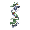
| ||||||||
|---|---|---|---|---|---|---|---|---|---|
| 1 | 
| ||||||||
| Unit cell |
|
-Components
| #1: Protein | Mass: 10842.916 Da / Num. of mol.: 2 / Fragment: UNP residues 416-509 Source method: isolated from a genetically manipulated source Source: (gene. exp.) Homo sapiens (human) / Gene: DVL2 / Plasmid: pETM-41 / Production host:  Escherichia coli BL21(DE3) (bacteria) / References: UniProt: O14641 Escherichia coli BL21(DE3) (bacteria) / References: UniProt: O14641#2: Water | ChemComp-HOH / | Water Mass: 18.015 Da / Num. of mol.: 82 / Source method: isolated from a natural source / Formula: H2O Mass: 18.015 Da / Num. of mol.: 82 / Source method: isolated from a natural source / Formula: H2O |
|---|
-Experimental details
-Experiment
| Experiment | Method: X-RAY DIFFRACTION / Number of used crystals: 1 |
|---|
- Sample preparation
| Crystal | Density Matthews: 2.45 Å3/Da / Density % sol: 49.78 % |
|---|---|
| Crystal grow | Temperature: 298 K / Method: vapor diffusion, sitting drop / pH: 6.5 / Details: 0.1 M MES, pH=6.5 0.2 M CaCl2 23% PEG350 MME |
-Data collection
| Diffraction | Mean temperature: 100 K |
|---|---|
| Diffraction source | Source: SYNCHROTRON / Site: Diamond  / Beamline: I04-1 / Wavelength: 0.92819 Å / Beamline: I04-1 / Wavelength: 0.92819 Å |
| Detector | Type: DECTRIS PILATUS 6M-F / Detector: PIXEL / Date: Oct 17, 2015 |
| Radiation | Protocol: SINGLE WAVELENGTH / Monochromatic (M) / Laue (L): M / Scattering type: x-ray |
| Radiation wavelength | Wavelength: 0.92819 Å / Relative weight: 1 |
| Reflection | Resolution: 1.84→32.7 Å / Num. obs: 17446 / % possible obs: 92 % / Redundancy: 13.7 % / Rmerge(I) obs: 0.107 / Net I/σ(I): 16.1 |
| Reflection shell | Resolution: 1.84→1.91 Å / Redundancy: 13.6 % / Rmerge(I) obs: 2.416 / Mean I/σ(I) obs: 0.98 / Num. unique all: 1733 |
- Processing
| Software |
| |||||||||||||||||||||||||||||||||||||||||||||||||||||||||||||||||||||||||||||||||||||||||||||||||||||||||||||||||||||||||||||||||||||||||||||||||||||||||||||||||||||||||||||||||||||||||||||||||||||||||||||||||||||||||||||||||
|---|---|---|---|---|---|---|---|---|---|---|---|---|---|---|---|---|---|---|---|---|---|---|---|---|---|---|---|---|---|---|---|---|---|---|---|---|---|---|---|---|---|---|---|---|---|---|---|---|---|---|---|---|---|---|---|---|---|---|---|---|---|---|---|---|---|---|---|---|---|---|---|---|---|---|---|---|---|---|---|---|---|---|---|---|---|---|---|---|---|---|---|---|---|---|---|---|---|---|---|---|---|---|---|---|---|---|---|---|---|---|---|---|---|---|---|---|---|---|---|---|---|---|---|---|---|---|---|---|---|---|---|---|---|---|---|---|---|---|---|---|---|---|---|---|---|---|---|---|---|---|---|---|---|---|---|---|---|---|---|---|---|---|---|---|---|---|---|---|---|---|---|---|---|---|---|---|---|---|---|---|---|---|---|---|---|---|---|---|---|---|---|---|---|---|---|---|---|---|---|---|---|---|---|---|---|---|---|---|---|---|---|---|---|---|---|---|---|---|---|---|---|---|---|---|---|---|
| Refinement | Method to determine structure: SAD / Resolution: 1.84→32.7 Å / Cor.coef. Fo:Fc: 0.959 / Cor.coef. Fo:Fc free: 0.948 / SU B: 11.932 / SU ML: 0.164 / Cross valid method: THROUGHOUT / σ(F): 0 / ESU R: 0.168 / ESU R Free: 0.158 / Stereochemistry target values: MAXIMUM LIKELIHOOD Details: HYDROGENS HAVE BEEN ADDED IN THE RIDING POSITIONS U VALUES : WITH TLS ADDED
| |||||||||||||||||||||||||||||||||||||||||||||||||||||||||||||||||||||||||||||||||||||||||||||||||||||||||||||||||||||||||||||||||||||||||||||||||||||||||||||||||||||||||||||||||||||||||||||||||||||||||||||||||||||||||||||||||
| Solvent computation | Ion probe radii: 0.8 Å / Shrinkage radii: 0.8 Å / VDW probe radii: 1.2 Å / Solvent model: MASK | |||||||||||||||||||||||||||||||||||||||||||||||||||||||||||||||||||||||||||||||||||||||||||||||||||||||||||||||||||||||||||||||||||||||||||||||||||||||||||||||||||||||||||||||||||||||||||||||||||||||||||||||||||||||||||||||||
| Displacement parameters | Biso max: 82.68 Å2 / Biso mean: 40.485 Å2 / Biso min: 25.09 Å2
| |||||||||||||||||||||||||||||||||||||||||||||||||||||||||||||||||||||||||||||||||||||||||||||||||||||||||||||||||||||||||||||||||||||||||||||||||||||||||||||||||||||||||||||||||||||||||||||||||||||||||||||||||||||||||||||||||
| Refinement step | Cycle: final / Resolution: 1.84→32.7 Å
| |||||||||||||||||||||||||||||||||||||||||||||||||||||||||||||||||||||||||||||||||||||||||||||||||||||||||||||||||||||||||||||||||||||||||||||||||||||||||||||||||||||||||||||||||||||||||||||||||||||||||||||||||||||||||||||||||
| Refine LS restraints |
| |||||||||||||||||||||||||||||||||||||||||||||||||||||||||||||||||||||||||||||||||||||||||||||||||||||||||||||||||||||||||||||||||||||||||||||||||||||||||||||||||||||||||||||||||||||||||||||||||||||||||||||||||||||||||||||||||
| LS refinement shell | Resolution: 1.844→1.891 Å / Total num. of bins used: 20
| |||||||||||||||||||||||||||||||||||||||||||||||||||||||||||||||||||||||||||||||||||||||||||||||||||||||||||||||||||||||||||||||||||||||||||||||||||||||||||||||||||||||||||||||||||||||||||||||||||||||||||||||||||||||||||||||||
| Refinement TLS params. | Method: refined / Refine-ID: X-RAY DIFFRACTION
| |||||||||||||||||||||||||||||||||||||||||||||||||||||||||||||||||||||||||||||||||||||||||||||||||||||||||||||||||||||||||||||||||||||||||||||||||||||||||||||||||||||||||||||||||||||||||||||||||||||||||||||||||||||||||||||||||
| Refinement TLS group |
|
