 Movie
Movie Controller

[English] 日本語
 Yorodumi
- PDB-5s8v: PanDDA analysis group deposition -- Crystal Structure of PHIP in ... -
+ Open data

- Basic information
| Entry | Database: PDB / ID: 5s8v | ||||||
|---|---|---|---|---|---|---|---|
| Title | PanDDA analysis group deposition -- Crystal Structure of PHIP in complex with Z198194396 synthetic derivative | ||||||
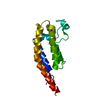
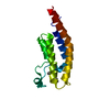
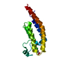
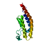
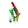
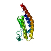
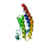
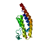
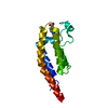
 Components Components | PH-interacting protein | ||||||
 Keywords Keywords |  SIGNALING PROTEIN / SGC - Diamond I04-1 fragment screening / PanDDA / XChemExplorer / Robotic chemistry / Crystal soaking / Reaction crudes SIGNALING PROTEIN / SGC - Diamond I04-1 fragment screening / PanDDA / XChemExplorer / Robotic chemistry / Crystal soaking / Reaction crudes | ||||||
| Function / homology |  Function and homology information Function and homology informationregulation of cell morphogenesis / positive regulation of insulin-like growth factor receptor signaling pathway / RHOBTB2 GTPase cycle / cytoskeleton organization / positive regulation of mitotic nuclear division / negative regulation of extrinsic apoptotic signaling pathway / regulation of protein phosphorylation / lysine-acetylated histone binding / insulin receptor binding / insulin receptor signaling pathway ...regulation of cell morphogenesis / positive regulation of insulin-like growth factor receptor signaling pathway / RHOBTB2 GTPase cycle / cytoskeleton organization / positive regulation of mitotic nuclear division / negative regulation of extrinsic apoptotic signaling pathway / regulation of protein phosphorylation / lysine-acetylated histone binding / insulin receptor binding / insulin receptor signaling pathway / regulation of cell shape / positive regulation of cell population proliferation / regulation of transcription by RNA polymerase II / negative regulation of apoptotic process / positive regulation of DNA-templated transcription / positive regulation of transcription by RNA polymerase II / nucleusSimilarity search - Function | ||||||
| Biological species |  Homo sapiens (human) Homo sapiens (human) | ||||||
| Method | X-RAY DIFFRACTION / SYNCHROTRON / FOURIER SYNTHESIS / molecular replacement / Resolution: 1.18 Å | ||||||
 Authors Authors | Grosjean, H. / Aimon, A. / Hassel-Hart , S. / Krojer, T. / Talon, R. / Douangamath, A. / Koekemoer, L. / Biggin, P.C. / Spencer, J. / von Delft, F. | ||||||
 Citation Citation | Journal: To Be Published Title: Crystal Structures of the second bromodomain of Pleckstrin homology domain interacting protein (PHIP) in space group C2 soaked with crude reaction mixtures Authors: Grosjean, H. / Aimon, A. / Hart , S. / Krojer, T. / Talon, R. / Douangamath, A. / Koekemoer, L. / Biggin, P.C. / Spencer, J. / von Delft, F. | ||||||
| History |
|
- Structure visualization
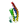
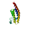
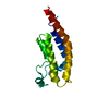
| Structure viewer | Molecule: MolmilJmol/JSmol |
|---|
- Downloads & links
-Download
| PDBx/mmCIF format | 5s8v.cif.gz | 44.2 KB | Display | PDBx/mmCIF format |
|---|---|---|---|---|
| PDB format | pdb5s8v.ent.gz | 29.8 KB | Display | PDB format |
| PDBx/mmJSON format | 5s8v.json.gz | Tree view | PDBx/mmJSON format | |
| Others |  Other downloads Other downloads |
-Validation report
| Arichive directory | https://data.pdbj.org/pub/pdb/validation_reports/s8/5s8vftp://data.pdbj.org/pub/pdb/validation_reports/s8/5s8v | HTTPS FTP |
|---|
-Group deposition
| ID | G_1002190 (26 entries) |
|---|---|
| Title | PanDDA analysis group deposition |
| Type | changed state |
| Description | XDomainX of XOrganismX PHIP screened against crude reaction mixtures by X-ray Crystallography at the XChem facility of Diamond Light Source beamline I04-1 |


-Related structure data
| Related structure data | 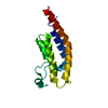 5rjiS S: Starting model for refinement |
|---|---|
| Similar structure data |


-Links
 PDBj
PDBj- Assembly
| Deposited unit | 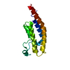
| |||||||||
|---|---|---|---|---|---|---|---|---|---|---|
| 1 |
| |||||||||
| Unit cell |
| |||||||||
| Components on special symmetry positions |
|
-Components
| #1: Protein | Mass: 17627.859 Da / Num. of mol.: 1 Source method: isolated from a genetically manipulated source Source: (gene. exp.) Homo sapiens (human) / Gene: PHIP, DCAF14, WDR11 / Production host:  Escherichia coli (E. coli) / References: UniProt: Q8WWQ0 Escherichia coli (E. coli) / References: UniProt: Q8WWQ0 |
|---|---|
| #2: Chemical | ChemComp-Y1A /   Mass: 265.308 Da / Num. of mol.: 1 / Source method: obtained synthetically / Formula: C13H19N3O3 Mass: 265.308 Da / Num. of mol.: 1 / Source method: obtained synthetically / Formula: C13H19N3O3 |
| #3: Water | ChemComp-HOH / Water Mass: 18.015 Da / Num. of mol.: 193 / Source method: isolated from a natural source / Formula: H2O Mass: 18.015 Da / Num. of mol.: 193 / Source method: isolated from a natural source / Formula: H2O |
-Experimental details
-Experiment
| Experiment | Method: X-RAY DIFFRACTION / Number of used crystals: 1 |
|---|
- Sample preparation
| Crystal | Density Matthews: 1.75 Å3/Da / Density % sol: 29.61 % |
|---|---|
| Crystal grow | Temperature: 277 K / Method: vapor diffusion, sitting drop / pH: 5.6 / Details: 20% PEG 8000, 0.04M POTASSIUM PHOSPHATE |
-Data collection
| Diffraction | Mean temperature: 100 K | |||||||||||||||||||||||||||
|---|---|---|---|---|---|---|---|---|---|---|---|---|---|---|---|---|---|---|---|---|---|---|---|---|---|---|---|---|
| Diffraction source | Source: SYNCHROTRON / Site: Diamond  / Beamline: I04-1 / Wavelength: 0.9159 Å / Beamline: I04-1 / Wavelength: 0.9159 Å | |||||||||||||||||||||||||||
| Detector | Type: DECTRIS PILATUS 6M / Detector: PIXEL / Date: Apr 11, 2019 | |||||||||||||||||||||||||||
| Radiation | Protocol: SINGLE WAVELENGTH / Scattering type: x-ray | |||||||||||||||||||||||||||
| Radiation wavelength | Wavelength: 0.9159 Å / Relative weight: 1 | |||||||||||||||||||||||||||
| Reflection | Resolution: 1.18→40.26 Å / Num. obs: 28785 / % possible obs: 88.7 % / Redundancy: 2.7 % / CC1/2: 0.999 / Rpim(I) all: 0.02 / Rrim(I) all: 0.036 / Net I/σ(I): 15 / Num. measured all: 76478 | |||||||||||||||||||||||||||
| Reflection shell | Diffraction-ID: 1
|
-Phasing
| Phasing | Method: molecular replacement |
|---|
- Processing
| Software |
| |||||||||||||||||||||||||||||||||||||||||||||||||||||||||||||||||||||||||||
|---|---|---|---|---|---|---|---|---|---|---|---|---|---|---|---|---|---|---|---|---|---|---|---|---|---|---|---|---|---|---|---|---|---|---|---|---|---|---|---|---|---|---|---|---|---|---|---|---|---|---|---|---|---|---|---|---|---|---|---|---|---|---|---|---|---|---|---|---|---|---|---|---|---|---|---|---|
| Refinement | Method to determine structure: FOURIER SYNTHESIS Starting model: 5RJI Resolution: 1.18→40.26 Å / Cor.coef. Fo:Fc: 0.969 / Cor.coef. Fo:Fc free: 0.954 / SU B: 1.08 / SU ML: 0.046 / Cross valid method: THROUGHOUT / σ(F): 0 / ESU R: 0.069 / ESU R Free: 0.07 / Stereochemistry target values: MAXIMUM LIKELIHOOD Details: HYDROGENS HAVE BEEN ADDED IN THE RIDING POSITIONS U VALUES : REFINED INDIVIDUALLY
| |||||||||||||||||||||||||||||||||||||||||||||||||||||||||||||||||||||||||||
| Solvent computation | Ion probe radii: 0.8 Å / Shrinkage radii: 0.8 Å / VDW probe radii: 1.2 Å / Solvent model: MASK | |||||||||||||||||||||||||||||||||||||||||||||||||||||||||||||||||||||||||||
| Displacement parameters | Biso max: 74.22 Å2 / Biso mean: 16.496 Å2 / Biso min: 7.29 Å2
| |||||||||||||||||||||||||||||||||||||||||||||||||||||||||||||||||||||||||||
| Refinement step | Cycle: final / Resolution: 1.18→40.26 Å
| |||||||||||||||||||||||||||||||||||||||||||||||||||||||||||||||||||||||||||
| Refine LS restraints |
| |||||||||||||||||||||||||||||||||||||||||||||||||||||||||||||||||||||||||||
| LS refinement shell | Resolution: 1.18→1.21 Å / Total num. of bins used: 20
|
