 Movie
Movie Controller

[English] 日本語
 Yorodumi
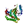
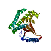
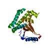
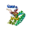
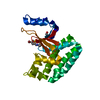
- PDB-5qiu: Covalent fragment group deposition -- Crystal Structure of OUTB2 ... -
+ Open data

- Basic information
| Entry | Database: PDB / ID: 5qiu | ||||||
|---|---|---|---|---|---|---|---|
| Title | Covalent fragment group deposition -- Crystal Structure of OUTB2 in complex with PCM-0103011 | ||||||
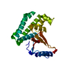
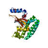
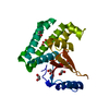
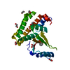
 Components Components | Ubiquitin thioesterase OTUB2 | ||||||
 Keywords Keywords | hydrolase/hydrolase inhibitor / SGC - Diamond I04-1 fragment screening / XChemExplorer / PROTEASE OTUB2 / DEUBIQUITINATING ENZYME OTUB2 / hydrolase-hydrolase inhibitor complex | ||||||
| Function / homology |  Function and homology information Function and homology informationnegative regulation of double-strand break repair / protein K11-linked deubiquitination / protein K48-linked deubiquitination / protein K63-linked deubiquitination / protein deubiquitination /  ubiquitin binding / Ovarian tumor domain proteases / ubiquitinyl hydrolase 1 / cysteine-type deubiquitinase activity / proteolysis / nucleus ubiquitin binding / Ovarian tumor domain proteases / ubiquitinyl hydrolase 1 / cysteine-type deubiquitinase activity / proteolysis / nucleusSimilarity search - Function | ||||||
| Biological species |  Homo sapiens (human) Homo sapiens (human) | ||||||
| Method | X-RAY DIFFRACTION / SYNCHROTRON / FOURIER SYNTHESIS / molecular replacement / Resolution: 1.56 Å | ||||||
 Authors Authors | Sethi, R. / Douangamath, A. / Resnick, E. / Bradley, A.R. / Collins, P. / Brandao-Neto, J. / Talon, R. / Krojer, T. / Bountra, C. / Arrowsmith, C.H. ...Sethi, R. / Douangamath, A. / Resnick, E. / Bradley, A.R. / Collins, P. / Brandao-Neto, J. / Talon, R. / Krojer, T. / Bountra, C. / Arrowsmith, C.H. / Edwards, A. / London, N. / von Delft, F. | ||||||
 Citation Citation | Journal: To Be Published Title: Covalent fragment group deposition Authors: Sethi, R. / Douangamath, A. / Resnick, E. / Bradley, A.R. / Collins, P. / Brandao-Neto, J. / Talon, R. / Krojer, T. / Bountra, C. / Arrowsmith, C.H. / Edwards, A. / London, N. / von Delft, F. | ||||||
| History |
|
- Structure visualization
| Structure viewer | Molecule: MolmilJmol/JSmol |
|---|
- Downloads & links
-Download
| PDBx/mmCIF format | 5qiu.cif.gz | 114 KB | Display | PDBx/mmCIF format |
|---|---|---|---|---|
| PDB format | pdb5qiu.ent.gz | 93.2 KB | Display | PDB format |
| PDBx/mmJSON format | 5qiu.json.gz | Tree view | PDBx/mmJSON format | |
| Others |  Other downloads Other downloads |
-Validation report
| Arichive directory | https://data.pdbj.org/pub/pdb/validation_reports/qi/5qiuftp://data.pdbj.org/pub/pdb/validation_reports/qi/5qiu | HTTPS FTP |
|---|
-Group deposition
| ID | G_1002053 (12 entries) |
|---|---|
| Title | Covalent fragment group deposition |
| Type | changed state |
| Description | human OUTB2 screened against covalent fragments by X-ray Crystallography at the XChem facility of Diamond Light Source beamline I04-1 |


-Related structure data
| Related structure data | 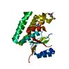 1tffS S: Starting model for refinement |
|---|---|
| Similar structure data |


-Links
 PDBj
PDBj

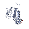
- Assembly
| Deposited unit | 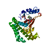
| ||||||||
|---|---|---|---|---|---|---|---|---|---|
| 1 |
| ||||||||
| Unit cell |
|
-Components
| #1: Protein | Mass: 26362.008 Da / Num. of mol.: 1 Source method: isolated from a genetically manipulated source Source: (gene. exp.) Homo sapiens (human) / Gene: OTUB2, C14orf137, OTB2, OTU2 / Production host:  Escherichia coli (E. coli) / References: UniProt: Q96DC9, ubiquitinyl hydrolase 1 Escherichia coli (E. coli) / References: UniProt: Q96DC9, ubiquitinyl hydrolase 1 | ||
|---|---|---|---|
| #2: Chemical | ChemComp-J5M /   Mass: 241.209 Da / Num. of mol.: 1 / Source method: obtained synthetically / Formula: C12H10F3NO Mass: 241.209 Da / Num. of mol.: 1 / Source method: obtained synthetically / Formula: C12H10F3NO | ||
| #3: Chemical | ChemComp-EDO / Ethylene glycol  Mass: 62.068 Da / Num. of mol.: 1 / Source method: obtained synthetically / Formula: C2H6O2 Mass: 62.068 Da / Num. of mol.: 1 / Source method: obtained synthetically / Formula: C2H6O2 | ||
| #4: Chemical | ChemComp-UNL / Num. of mol.: 5 / Source method: obtained synthetically #5: Water | ChemComp-HOH / | Water Mass: 18.015 Da / Num. of mol.: 234 / Source method: isolated from a natural source / Formula: H2O Mass: 18.015 Da / Num. of mol.: 234 / Source method: isolated from a natural source / Formula: H2O |
-Experimental details
-Experiment
| Experiment | Method: X-RAY DIFFRACTION / Number of used crystals: 1 |
|---|
- Sample preparation
| Crystal | Density Matthews: 2.36 Å3/Da / Density % sol: 47.96 % / Mosaicity: 0.12 ° |
|---|---|
| Crystal grow | Temperature: 293 K / Method: vapor diffusion, sitting drop / pH: 7 Details: 16% PEG4K, 0.1M HEPES pH 7.0, 8% 2-propanol, 5 mM DTT |
-Data collection
| Diffraction | Mean temperature: 100 K | ||||||||||||||||||||||||||||||
|---|---|---|---|---|---|---|---|---|---|---|---|---|---|---|---|---|---|---|---|---|---|---|---|---|---|---|---|---|---|---|---|
| Diffraction source | Source: SYNCHROTRON / Site: Diamond  / Beamline: I04-1 / Wavelength: 0.92819 Å / Beamline: I04-1 / Wavelength: 0.92819 Å | ||||||||||||||||||||||||||||||
| Detector | Type: DECTRIS PILATUS 6M / Detector: PIXEL / Date: Jun 28, 2017 | ||||||||||||||||||||||||||||||
| Radiation | Protocol: SINGLE WAVELENGTH / Scattering type: x-ray | ||||||||||||||||||||||||||||||
| Radiation wavelength | Wavelength: 0.92819 Å / Relative weight: 1 | ||||||||||||||||||||||||||||||
| Reflection | Resolution: 1.56→29.21 Å / Num. obs: 34960 / % possible obs: 99.8 % / Redundancy: 3.4 % / CC1/2: 0.998 / Rmerge(I) obs: 0.078 / Rpim(I) all: 0.049 / Rrim(I) all: 0.092 / Net I/σ(I): 9.9 / Num. measured all: 117970 / Scaling rejects: 0 | ||||||||||||||||||||||||||||||
| Reflection shell | Diffraction-ID: 1
|
-Phasing
| Phasing | Method: molecular replacement |
|---|
- Processing
| Software |
| ||||||||||||||||||||||||||||||||||||||||||||||||||||||||||||||||||||||||||||||||||||||||||
|---|---|---|---|---|---|---|---|---|---|---|---|---|---|---|---|---|---|---|---|---|---|---|---|---|---|---|---|---|---|---|---|---|---|---|---|---|---|---|---|---|---|---|---|---|---|---|---|---|---|---|---|---|---|---|---|---|---|---|---|---|---|---|---|---|---|---|---|---|---|---|---|---|---|---|---|---|---|---|---|---|---|---|---|---|---|---|---|---|---|---|---|
| Refinement | Method to determine structure: FOURIER SYNTHESIS Starting model: 1TFF Resolution: 1.56→29.23 Å / Cor.coef. Fo:Fc: 0.975 / Cor.coef. Fo:Fc free: 0.953 / SU B: 4.676 / SU ML: 0.072 / Cross valid method: THROUGHOUT / σ(F): 0 / ESU R: 0.088 / ESU R Free: 0.084 / Stereochemistry target values: MAXIMUM LIKELIHOOD Details: HYDROGENS HAVE BEEN ADDED IN THE RIDING POSITIONS U VALUES : REFINED INDIVIDUALLY
| ||||||||||||||||||||||||||||||||||||||||||||||||||||||||||||||||||||||||||||||||||||||||||
| Solvent computation | Ion probe radii: 0.8 Å / Shrinkage radii: 0.8 Å / VDW probe radii: 1.2 Å / Solvent model: MASK | ||||||||||||||||||||||||||||||||||||||||||||||||||||||||||||||||||||||||||||||||||||||||||
| Displacement parameters | Biso max: 91.86 Å2 / Biso mean: 21.006 Å2 / Biso min: 10.04 Å2
| ||||||||||||||||||||||||||||||||||||||||||||||||||||||||||||||||||||||||||||||||||||||||||
| Refinement step | Cycle: final / Resolution: 1.56→29.23 Å
| ||||||||||||||||||||||||||||||||||||||||||||||||||||||||||||||||||||||||||||||||||||||||||
| Refine LS restraints |
| ||||||||||||||||||||||||||||||||||||||||||||||||||||||||||||||||||||||||||||||||||||||||||
| LS refinement shell | Resolution: 1.561→1.601 Å / Total num. of bins used: 20
|
