 Movie
Movie Controller

[English] 日本語
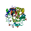
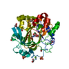
 Yorodumi
- PDB-5ph6: PanDDA analysis group deposition -- Crystal Structure of JMJD2D i... -
+ Open data

- Basic information
| Entry | Database: PDB / ID: 5ph6 | ||||||
|---|---|---|---|---|---|---|---|
| Title | PanDDA analysis group deposition -- Crystal Structure of JMJD2D in complex with N09736a | ||||||
 Components Components | Lysine-specific demethylase 4D | ||||||
 Keywords Keywords |  OXIDOREDUCTASE / PanDDA / SGC - Diamond I04-1 fragment screening / Jmj domain / epigenetics OXIDOREDUCTASE / PanDDA / SGC - Diamond I04-1 fragment screening / Jmj domain / epigenetics | ||||||
| Function / homology |  Function and homology information Function and homology informationpositive regulation of chromatin binding / histone H3K9me2/H3K9me3 demethylase activity / [histone H3]-trimethyl-L-lysine9 demethylase / positive regulation of double-strand break repair via nonhomologous end joining / histone H3K9 demethylase activity / histone demethylase activity / pericentric heterochromatin / cellular response to ionizing radiation / regulation of protein phosphorylation / double-strand break repair via homologous recombination ...positive regulation of chromatin binding / histone H3K9me2/H3K9me3 demethylase activity / [histone H3]-trimethyl-L-lysine9 demethylase / positive regulation of double-strand break repair via nonhomologous end joining / histone H3K9 demethylase activity / histone demethylase activity / pericentric heterochromatin / cellular response to ionizing radiation / regulation of protein phosphorylation / double-strand break repair via homologous recombination / HDMs demethylate histones / chromatin DNA binding / site of double-strand break / regulation of gene expression / blood microparticle / damaged DNA binding / chromatin remodeling / inflammatory response / chromatin / nucleoplasm / metal ion binding / nucleusSimilarity search - Function | ||||||
| Biological species |  Homo sapiens (human) Homo sapiens (human) | ||||||
| Method | X-RAY DIFFRACTION / SYNCHROTRON / FOURIER SYNTHESIS / molecular replacement / Resolution: 1.742 Å | ||||||
 Authors Authors | Pearce, N.M. / Krojer, T. / Talon, R. / Bradley, A.R. / Fairhead, M. / Sethi, R. / Wright, N. / MacLean, E. / Collins, P. / Brandao-Neto, J. ...Pearce, N.M. / Krojer, T. / Talon, R. / Bradley, A.R. / Fairhead, M. / Sethi, R. / Wright, N. / MacLean, E. / Collins, P. / Brandao-Neto, J. / Douangamath, A. / Renjie, Z. / Dias, A. / Vollmar, M. / Ng, J. / Szykowska, A. / Burgess-Brown, N. / Brennan, P.E. / Cox, O. / Oppermann, U. / Bountra, C. / Arrowsmith, C.H. / Edwards, A. / von Delft, F. | ||||||
 Citation Citation | Journal: Nat Commun / Year: 2017 Title: A multi-crystal method for extracting obscured crystallographic states from conventionally uninterpretable electron density. Authors: Pearce, N.M. / Krojer, T. / Bradley, A.R. / Collins, P. / Nowak, R.P. / Talon, R. / Marsden, B.D. / Kelm, S. / Shi, J. / Deane, C.M. / von Delft, F. | ||||||
| History |
|
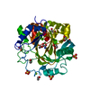
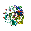
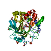
- Structure visualization
| Structure viewer | Molecule: MolmilJmol/JSmol |
|---|
- Downloads & links
-Download
| PDBx/mmCIF format | 5ph6.cif.gz | 105.1 KB | Display | PDBx/mmCIF format |
|---|---|---|---|---|
| PDB format | pdb5ph6.ent.gz | 78.1 KB | Display | PDB format |
| PDBx/mmJSON format | 5ph6.json.gz | Tree view | PDBx/mmJSON format | |
| Others |  Other downloads Other downloads |
-Validation report
| Arichive directory | https://data.pdbj.org/pub/pdb/validation_reports/ph/5ph6ftp://data.pdbj.org/pub/pdb/validation_reports/ph/5ph6 | HTTPS FTP |
|---|
-Group deposition
| ID | G_1002020 (24 entries) |
|---|---|
| Title | PanDDA analysis group deposition of models with modelled events (e.g. bound ligands) |
| Type | changed state |
| Description | Jmjc domain of human JMJD2D screened against the ZENOBIA Fragment Library by X-ray Crystallography. Check out the PanDDA event maps at https://zenodo.org/record/290220/files/0_index.html |


-Related structure data
| Related structure data | 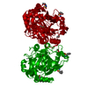 4d6rS S: Starting model for refinement |
|---|---|
| Similar structure data |


-Links
 PDBj
PDBj
- Assembly
| Deposited unit | 
| ||||||||
|---|---|---|---|---|---|---|---|---|---|
| 1 |
| ||||||||
| Unit cell |
| ||||||||
| Components on special symmetry positions |
|
-Components
-Protein , 1 types, 1 molecules A
| #1: Protein | Mass: 42050.539 Da / Num. of mol.: 1 Source method: isolated from a genetically manipulated source Source: (gene. exp.) Homo sapiens (human) / Gene: KDM4D, JHDM3D, JMJD2D / Production host:  escherichia coli (E. coli) / References: UniProt: Q6B0I6 escherichia coli (E. coli) / References: UniProt: Q6B0I6 |
|---|
-Non-polymers , 7 types, 396 molecules 


| #2: Chemical | ChemComp-ZN /  Mass: 65.409 Da / Num. of mol.: 1 / Source method: obtained synthetically / Formula: Zn Mass: 65.409 Da / Num. of mol.: 1 / Source method: obtained synthetically / Formula: Zn | ||||||
|---|---|---|---|---|---|---|---|
| #3: Chemical | ChemComp-NI / Nickel Mass: 58.693 Da / Num. of mol.: 1 / Source method: obtained synthetically / Formula: Ni Mass: 58.693 Da / Num. of mol.: 1 / Source method: obtained synthetically / Formula: Ni | ||||||
| #4: Chemical | ChemComp-OGA / N-Oxalylglycine Mass: 147.086 Da / Num. of mol.: 1 / Source method: obtained synthetically / Formula: C4H5NO5 / Comment: inhibitor*YM Mass: 147.086 Da / Num. of mol.: 1 / Source method: obtained synthetically / Formula: C4H5NO5 / Comment: inhibitor*YM | ||||||
| #5: Chemical | ChemComp-EDO / Ethylene glycol Mass: 62.068 Da / Num. of mol.: 8 / Source method: obtained synthetically / Formula: C2H6O2 Mass: 62.068 Da / Num. of mol.: 8 / Source method: obtained synthetically / Formula: C2H6O2#6: Chemical | Sulfate Mass: 96.063 Da / Num. of mol.: 3 / Source method: obtained synthetically / Formula: SO4 Mass: 96.063 Da / Num. of mol.: 3 / Source method: obtained synthetically / Formula: SO4#7: Chemical | Isatin Mass: 147.131 Da / Num. of mol.: 2 / Source method: obtained synthetically / Formula: C8H5NO2 Mass: 147.131 Da / Num. of mol.: 2 / Source method: obtained synthetically / Formula: C8H5NO2#8: Water | ChemComp-HOH / | WaterMass: 18.015 Da / Num. of mol.: 380 / Source method: isolated from a natural source / Formula: H2O |
-Details
| Nonpolymer details | yes c1ccc2c(c1)C(C(N2)=O)=O None -14 -16 -16 c1ccc2c(c1)C(C(N2)=O)=O 4 - High Confidence None 0. ... |
|---|
-Experimental details
-Experiment
| Experiment | Method: X-RAY DIFFRACTION / Number of used crystals: 1 |
|---|
- Sample preparation
| Crystal | Density Matthews: 2.26 Å3/Da / Density % sol: 45.64 % / Mosaicity: 0.05 ° |
|---|---|
| Crystal grow | Temperature: 298 K / Method: vapor diffusion, sitting drop / pH: 7 Details: 28% PEG3350 -- 0.1M HEPES pH 7.0 -- 0.25M ammonium sulfate |
-Data collection
| Diffraction | Mean temperature: 100 K | ||||||||||||||||||||||||||||||
|---|---|---|---|---|---|---|---|---|---|---|---|---|---|---|---|---|---|---|---|---|---|---|---|---|---|---|---|---|---|---|---|
| Diffraction source | Source: SYNCHROTRON / Site: Diamond  / Beamline: I03 / Wavelength: 0.9763 Å / Beamline: I03 / Wavelength: 0.9763 Å | ||||||||||||||||||||||||||||||
| Detector | Type: DECTRIS PILATUS 6M / Detector: PIXEL / Date: Dec 15, 2012 | ||||||||||||||||||||||||||||||
| Radiation | Protocol: SINGLE WAVELENGTH / Scattering type: x-ray | ||||||||||||||||||||||||||||||
| Radiation wavelength | Wavelength: 0.9763 Å / Relative weight: 1 | ||||||||||||||||||||||||||||||
| Reflection | Resolution: 1.74→29.316 Å / Num. obs: 40370 / % possible obs: 99.9 % / Redundancy: 13 % / Biso Wilson estimate: 16.71 Å2 / CC1/2: 0.996 / Rmerge(I) obs: 0.135 / Rpim(I) all: 0.039 / Rrim(I) all: 0.141 / Net I/σ(I): 13.5 / Num. measured all: 523536 / Scaling rejects: 0 | ||||||||||||||||||||||||||||||
| Reflection shell | Diffraction-ID: 1
|
-Phasing
| Phasing | Method: molecular replacement |
|---|
- Processing
| Software |
| |||||||||||||||||||||||||||||||||||||||||||||||||||||||||||||||||||||||||||||||||||||||||||||||||||||||||
|---|---|---|---|---|---|---|---|---|---|---|---|---|---|---|---|---|---|---|---|---|---|---|---|---|---|---|---|---|---|---|---|---|---|---|---|---|---|---|---|---|---|---|---|---|---|---|---|---|---|---|---|---|---|---|---|---|---|---|---|---|---|---|---|---|---|---|---|---|---|---|---|---|---|---|---|---|---|---|---|---|---|---|---|---|---|---|---|---|---|---|---|---|---|---|---|---|---|---|---|---|---|---|---|---|---|---|
| Refinement | Method to determine structure: FOURIER SYNTHESIS Starting model: 4D6R Resolution: 1.742→29.316 Å / SU ML: 0.15 / Cross valid method: THROUGHOUT / σ(F): 1.34 / Phase error: 17.49 / Stereochemistry target values: ML
| |||||||||||||||||||||||||||||||||||||||||||||||||||||||||||||||||||||||||||||||||||||||||||||||||||||||||
| Solvent computation | Shrinkage radii: 0.9 Å / VDW probe radii: 1.11 Å / Solvent model: FLAT BULK SOLVENT MODEL | |||||||||||||||||||||||||||||||||||||||||||||||||||||||||||||||||||||||||||||||||||||||||||||||||||||||||
| Displacement parameters | Biso max: 124.5 Å2 / Biso mean: 20.1731 Å2 / Biso min: 6.38 Å2 | |||||||||||||||||||||||||||||||||||||||||||||||||||||||||||||||||||||||||||||||||||||||||||||||||||||||||
| Refinement step | Cycle: final / Resolution: 1.742→29.316 Å
| |||||||||||||||||||||||||||||||||||||||||||||||||||||||||||||||||||||||||||||||||||||||||||||||||||||||||
| Refine LS restraints |
| |||||||||||||||||||||||||||||||||||||||||||||||||||||||||||||||||||||||||||||||||||||||||||||||||||||||||
| LS refinement shell | Refine-ID: X-RAY DIFFRACTION / Total num. of bins used: 14
|
