 Movie
Movie Controller

[English] 日本語
 Yorodumi
- PDB-5oj6: Crystal structure of the chicken MDGA1 ectodomain in complex with... -
+ Open data

- Basic information
| Entry | Database: PDB / ID: 5oj6 | ||||||||||||||||||||||||||||||||||||||||||
|---|---|---|---|---|---|---|---|---|---|---|---|---|---|---|---|---|---|---|---|---|---|---|---|---|---|---|---|---|---|---|---|---|---|---|---|---|---|---|---|---|---|---|---|
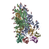
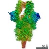
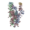
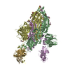
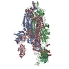
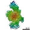
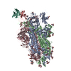
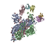
| Title | Crystal structure of the chicken MDGA1 ectodomain in complex with the human neuroligin 1 (NL1(-A-B)) cholinesterase domain. | ||||||||||||||||||||||||||||||||||||||||||
 Components Components |
| ||||||||||||||||||||||||||||||||||||||||||
 Keywords Keywords |  CELL ADHESION / neuroligin-neurexin / synapse formation / austism spectrum disorders (ASDs) / synaptic transmission CELL ADHESION / neuroligin-neurexin / synapse formation / austism spectrum disorders (ASDs) / synaptic transmission | ||||||||||||||||||||||||||||||||||||||||||
| Function / homology |  Function and homology information Function and homology informationneurexin clustering involved in presynaptic membrane assembly / asymmetric, glutamatergic, excitatory synapse / cytoskeletal matrix organization at active zone / cell-cell adhesion involved in synapse maturation / positive regulation of circadian sleep/wake cycle, wakefulness / protein complex involved in cell-cell adhesion / positive regulation of neuromuscular synaptic transmission / terminal button organization / neuron to neuron synapse / positive regulation of synaptic vesicle exocytosis ...neurexin clustering involved in presynaptic membrane assembly / asymmetric, glutamatergic, excitatory synapse / cytoskeletal matrix organization at active zone / cell-cell adhesion involved in synapse maturation / positive regulation of circadian sleep/wake cycle, wakefulness / protein complex involved in cell-cell adhesion / positive regulation of neuromuscular synaptic transmission / terminal button organization / neuron to neuron synapse / positive regulation of synaptic vesicle exocytosis / neuronal signal transduction / excitatory synapse assembly / postsynaptic density protein 95 clustering / negative regulation of dendritic spine morphogenesis / postsynaptic membrane assembly / synapse maturation / presynaptic membrane assembly / maintenance of synapse structure / synaptic vesicle targeting / inhibitory synapse / synaptic vesicle clustering / neuron cell-cell adhesion / presynapse assembly / neurexin family protein binding / receptor localization to synapse / filopodium tip / protein localization to synapse / NMDA glutamate receptor clustering / neuron projection arborization / positive regulation of synaptic vesicle endocytosis / calcium-dependent cell-cell adhesion via plasma membrane cell adhesion molecules / regulation of AMPA receptor activity / AMPA glutamate receptor clustering / postsynaptic specialization membrane / Neurexins and neuroligins / positive regulation of synapse assembly / positive regulation of ruffle assembly / regulation of NMDA receptor activity / positive regulation of intracellular signal transduction / positive regulation of filopodium assembly / heterophilic cell-cell adhesion via plasma membrane cell adhesion molecules / regulation of neuron differentiation / positive regulation of dendritic spine development / positive regulation of excitatory postsynaptic potential / synaptic vesicle endocytosis / excitatory synapse / GABA-ergic synapse / protein targeting / synaptic cleft / cell adhesion molecule binding / synapse assembly / cellular response to calcium ion / positive regulation of synaptic transmission, glutamatergic / neuron projection morphogenesis / PDZ domain binding / positive regulation of synaptic transmission, GABAergic / neuron migration / modulation of chemical synaptic transmission / neuromuscular junction / establishment of protein localization / positive regulation of neuron projection development / neuron projection development / rhythmic process / presynapse / signaling receptor activity / nervous system development / amyloid-beta binding / scaffold protein binding / postsynapse / chemical synaptic transmission / postsynaptic membrane / dendritic spine / postsynaptic density / receptor complex / glutamatergic synapse / synapse / dendrite / Golgi apparatus / cell surface / membrane / identical protein binding / plasma membraneSimilarity search - Function | ||||||||||||||||||||||||||||||||||||||||||
| Biological species |  Homo sapiens (human) Homo sapiens (human) Gallus gallus (chicken) Gallus gallus (chicken) | ||||||||||||||||||||||||||||||||||||||||||
| Method | X-RAY DIFFRACTION / SYNCHROTRON / MOLECULAR REPLACEMENT / Resolution: 3.3 Å | ||||||||||||||||||||||||||||||||||||||||||
 Authors Authors | Elegheert, J. / Clayton, A.J. / Aricescu, A.R. | ||||||||||||||||||||||||||||||||||||||||||
| Funding support |  United Kingdom, United Kingdom,  Canada, Canada,  United States, 13items United States, 13items
| ||||||||||||||||||||||||||||||||||||||||||
 Citation Citation | Journal: Neuron / Year: 2017 Title: Structural Mechanism for Modulation of Synaptic Neuroligin-Neurexin Signaling by MDGA Proteins. Authors: Elegheert, J. / Cvetkovska, V. / Clayton, A.J. / Heroven, C. / Vennekens, K.M. / Smukowski, S.N. / Regan, M.C. / Jia, W. / Smith, A.C. / Furukawa, H. / Savas, J.N. / de Wit, J. / Begbie, J. ...Authors: Elegheert, J. / Cvetkovska, V. / Clayton, A.J. / Heroven, C. / Vennekens, K.M. / Smukowski, S.N. / Regan, M.C. / Jia, W. / Smith, A.C. / Furukawa, H. / Savas, J.N. / de Wit, J. / Begbie, J. / Craig, A.M. / Aricescu, A.R. | ||||||||||||||||||||||||||||||||||||||||||
| History |
|
- Structure visualization
| Structure viewer | Molecule: MolmilJmol/JSmol |
|---|
- Downloads & links
-Download
| PDBx/mmCIF format | 5oj6.cif.gz | 269.6 KB | Display | PDBx/mmCIF format |
|---|---|---|---|---|
| PDB format | pdb5oj6.ent.gz | 208.4 KB | Display | PDB format |
| PDBx/mmJSON format | 5oj6.json.gz | Tree view | PDBx/mmJSON format | |
| Others |  Other downloads Other downloads |
-Validation report
| Arichive directory | https://data.pdbj.org/pub/pdb/validation_reports/oj/5oj6ftp://data.pdbj.org/pub/pdb/validation_reports/oj/5oj6 | HTTPS FTP |
|---|
-Related structure data
| Related structure data |  5oj2SC  5ojkC S: Starting model for refinement C: citing same article ( |
|---|---|
| Similar structure data |
-Links
 PDBj
PDBj


- Assembly
| Deposited unit | 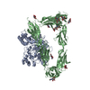
| ||||||||
|---|---|---|---|---|---|---|---|---|---|
| 1 | 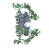
| ||||||||
| Unit cell |
|
-Components
| #1: Protein | Mass: 64397.254 Da / Num. of mol.: 1 Source method: isolated from a genetically manipulated source Source: (gene. exp.) Homo sapiens (human) / Gene: NLGN1, KIAA1070 / Plasmid: pHLsec / Cell line (production host): HEK293S GnTI-/- / Production host: Homo sapiens (human) / References: UniProt: Q8N2Q7 |
|---|---|
| #2: Protein | Mass: 102143.867 Da / Num. of mol.: 1 Source method: isolated from a genetically manipulated source Source: (gene. exp.) Gallus gallus (chicken) / Gene: MDGA1 / Plasmid: pHLsec / Cell line (production host): HEK293S GnTI-/- / Production host: Homo sapiens (human) / References: UniProt: Q0WYX8 |
| #3: Polysaccharide | beta-D-mannopyranose-(1-4)-2-acetamido-2-deoxy-beta-D-glucopyranose-(1-4)-2-acetamido-2-deoxy-beta- ...beta-D-mannopyranose-(1-4)-2-acetamido-2-deoxy-beta-D-glucopyranose-(1-4)-2-acetamido-2-deoxy-beta-D-glucopyranose / Mass: 586.542 Da / Num. of mol.: 1 Source method: isolated from a genetically manipulated source |
| #4: Sugar | ChemComp-NAG / N-Acetylglucosamine  Type: D-saccharide, beta linking / Mass: 221.208 Da / Num. of mol.: 8 Type: D-saccharide, beta linking / Mass: 221.208 Da / Num. of mol.: 8Source method: isolated from a genetically manipulated source Formula: C8H15NO6 |
-Experimental details
-Experiment
| Experiment | Method: X-RAY DIFFRACTION / Number of used crystals: 1 |
|---|
- Sample preparation
| Crystal | Density Matthews: 2.92 Å3/Da / Density % sol: 57.84 % |
|---|---|
| Crystal grow | Temperature: 293 K / Method: vapor diffusion, sitting drop / Details: 0.1M Na.HEPES pH 7.0, 7.5% w/v PEG8000 |
-Data collection
| Diffraction | Mean temperature: 80 K |
|---|---|
| Diffraction source | Source: SYNCHROTRON / Site: Diamond / Beamline: I04-1 / Wavelength: 0.92 Å |
| Detector | Type: DECTRIS PILATUS 6M / Detector: PIXEL / Date: May 15, 2014 |
| Radiation | Protocol: SINGLE WAVELENGTH / Monochromatic (M) / Laue (L): M / Scattering type: x-ray |
| Radiation wavelength | Wavelength: 0.92 Å / Relative weight: 1 |
| Reflection | Resolution: 3.3→92.07 Å / Num. obs: 29576 / % possible obs: 98.7 % / Redundancy: 4.5 % / CC1/2: 0.999 / Net I/σ(I): 15.6 |
| Reflection shell | Resolution: 3.3→3.39 Å / Redundancy: 4.6 % / Mean I/σ(I) obs: 1.6 / Num. unique obs: 2080 / CC1/2: 0.638 / Rrim(I) all: 1.063 / % possible all: 95.4 |
- Processing
| Software |
| ||||||||||||||||||||||||||||||||||||||||||||||||||||||||||||||||||||||||||||||||||||
|---|---|---|---|---|---|---|---|---|---|---|---|---|---|---|---|---|---|---|---|---|---|---|---|---|---|---|---|---|---|---|---|---|---|---|---|---|---|---|---|---|---|---|---|---|---|---|---|---|---|---|---|---|---|---|---|---|---|---|---|---|---|---|---|---|---|---|---|---|---|---|---|---|---|---|---|---|---|---|---|---|---|---|---|---|---|
| Refinement | Method to determine structure: MOLECULAR REPLACEMENT Starting model: 5OJ2 Resolution: 3.3→72.362 Å / SU ML: 0.47 / Cross valid method: FREE R-VALUE / σ(F): 1.34 / Phase error: 28.59
| ||||||||||||||||||||||||||||||||||||||||||||||||||||||||||||||||||||||||||||||||||||
| Solvent computation | Shrinkage radii: 0.8 Å / VDW probe radii: 1 Å | ||||||||||||||||||||||||||||||||||||||||||||||||||||||||||||||||||||||||||||||||||||
| Refinement step | Cycle: LAST / Resolution: 3.3→72.362 Å
| ||||||||||||||||||||||||||||||||||||||||||||||||||||||||||||||||||||||||||||||||||||
| Refine LS restraints |
| ||||||||||||||||||||||||||||||||||||||||||||||||||||||||||||||||||||||||||||||||||||
| LS refinement shell |
|
