 Movie
Movie Controller


+ Open data

- Basic information
| Entry | Database: PDB / ID: 5oi7 | ||||||
|---|---|---|---|---|---|---|---|
| Title | Human CEP85 - coiled coil domain 4 | ||||||
 Components Components | Centrosomal protein of 85 kDa Centrosome Centrosome | ||||||
 Keywords Keywords | PROTEIN BINDING / Centrosomes / centriole / CEP85 / STIL | ||||||
| Function / homology |  Function and homology information Function and homology informationregulation of mitotic centrosome separation / pericentriolar material / chromosome segregation / negative regulation of protein kinase activity / spindle pole / centrosome / nucleolus / Golgi apparatus / cytosolSimilarity search - Function | ||||||
| Biological species |  Homo sapiens (human) Homo sapiens (human) | ||||||
| Method | X-RAY DIFFRACTION / MOLECULAR REPLACEMENT / Resolution: 1.67 Å | ||||||
 Authors Authors | van Breugel, M. | ||||||
| Funding support |  United Kingdom, 1items United Kingdom, 1items
| ||||||
 Citation Citation | Journal: Nat Commun / Year: 2018 Title: Direct binding of CEP85 to STIL ensures robust PLK4 activation and efficient centriole assembly. Authors: Liu, Y. / Gupta, G.D. / Barnabas, D.D. / Agircan, F.G. / Mehmood, S. / Wu, D. / Coyaud, E. / Johnson, C.M. / McLaughlin, S.H. / Andreeva, A. / Freund, S.M.V. / Robinson, C.V. / Cheung, S.W.T. ...Authors: Liu, Y. / Gupta, G.D. / Barnabas, D.D. / Agircan, F.G. / Mehmood, S. / Wu, D. / Coyaud, E. / Johnson, C.M. / McLaughlin, S.H. / Andreeva, A. / Freund, S.M.V. / Robinson, C.V. / Cheung, S.W.T. / Raught, B. / Pelletier, L. / van Breugel, M. | ||||||
| History |
|
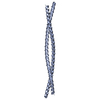
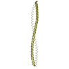
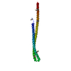
- Structure visualization
| Structure viewer | Molecule: MolmilJmol/JSmol |
|---|
- Downloads & links
-Download
| PDBx/mmCIF format | 5oi7.cif.gz | 54.8 KB | Display | PDBx/mmCIF format |
|---|---|---|---|---|
| PDB format | pdb5oi7.ent.gz | 39.1 KB | Display | PDB format |
| PDBx/mmJSON format | 5oi7.json.gz | Tree view | PDBx/mmJSON format | |
| Others |  Other downloads Other downloads |
-Validation report
| Arichive directory | https://data.pdbj.org/pub/pdb/validation_reports/oi/5oi7ftp://data.pdbj.org/pub/pdb/validation_reports/oi/5oi7 | HTTPS FTP |
|---|
-Related structure data
| Related structure data |  5oi9C  5oidC  2q6qS S: Starting model for refinement C: citing same article ( |
|---|---|
| Similar structure data |


-Links
 PDBj
PDBj- Assembly
| Deposited unit | 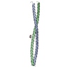
| ||||||||
|---|---|---|---|---|---|---|---|---|---|
| 1 |
| ||||||||
| Unit cell |
|
-Components
| #1: Protein | Centrosome / Cep85 / Coiled-coil domain-containing protein 21 Mass: 10347.432 Da / Num. of mol.: 2 Source method: isolated from a genetically manipulated source Source: (gene. exp.) Homo sapiens (human) / Gene: CEP85, CCDC21 / Production host:  Escherichia coli (E. coli) / Variant (production host): C41 / References: UniProt: Q6P2H3 Escherichia coli (E. coli) / Variant (production host): C41 / References: UniProt: Q6P2H3#2: Chemical | ChemComp-MPD / ( | 2-Methyl-2,4-pentanediol  Mass: 118.174 Da / Num. of mol.: 1 / Source method: obtained synthetically / Formula: C6H14O2 / Comment: precipitant*YM Mass: 118.174 Da / Num. of mol.: 1 / Source method: obtained synthetically / Formula: C6H14O2 / Comment: precipitant*YM#3: Water | ChemComp-HOH / | Water Mass: 18.015 Da / Num. of mol.: 227 / Source method: isolated from a natural source / Formula: H2O Mass: 18.015 Da / Num. of mol.: 227 / Source method: isolated from a natural source / Formula: H2O |
|---|
-Experimental details
-Experiment
| Experiment | Method: X-RAY DIFFRACTION / Number of used crystals: 1 |
|---|
- Sample preparation
| Crystal | Density Matthews: 2.17 Å3/Da / Density % sol: 43.29 % |
|---|---|
| Crystal grow | Temperature: 292 K / Method: vapor diffusion, sitting drop Details: 100 mM Na-HEPES, pH 7.7 200 mM Na-Citrate 36 % (v/v) MPD |
-Data collection
| Diffraction | Mean temperature: 100 K |
|---|---|
| Diffraction source | Source: ROTATING ANODE / Type: RIGAKU FR-E+ SUPERBRIGHT / Wavelength: 1.54179 Å |
| Detector | Type: MARRESEARCH / Detector: IMAGE PLATE / Date: Oct 22, 2014 |
| Radiation | Protocol: SINGLE WAVELENGTH / Monochromatic (M) / Laue (L): M / Scattering type: x-ray |
| Radiation wavelength | Wavelength: 1.54179 Å / Relative weight: 1 |
| Reflection | Resolution: 1.67→36.98 Å / Num. obs: 21341 / % possible obs: 98.9 % / Redundancy: 11.1 % / Rmerge(I) obs: 0.202 / Rpim(I) all: 0.063 / Net I/σ(I): 8.6 |
| Reflection shell | Resolution: 1.67→1.76 Å / Redundancy: 10.1 % / Rmerge(I) obs: 1.552 / Mean I/σ(I) obs: 1.4 / Num. unique obs: 2852 / CC1/2: 0.474 / Rpim(I) all: 0.506 / % possible all: 92.2 |
- Processing
| Software |
| |||||||||||||||||||||||||||||||||||||||||||||||||||||||||||||||||||||||||||||||||||||||||||||||||||||||||
|---|---|---|---|---|---|---|---|---|---|---|---|---|---|---|---|---|---|---|---|---|---|---|---|---|---|---|---|---|---|---|---|---|---|---|---|---|---|---|---|---|---|---|---|---|---|---|---|---|---|---|---|---|---|---|---|---|---|---|---|---|---|---|---|---|---|---|---|---|---|---|---|---|---|---|---|---|---|---|---|---|---|---|---|---|---|---|---|---|---|---|---|---|---|---|---|---|---|---|---|---|---|---|---|---|---|---|
| Refinement | Method to determine structure: MOLECULAR REPLACEMENT Starting model: 2Q6Q Resolution: 1.67→36.976 Å / SU ML: 0.23 / Cross valid method: FREE R-VALUE / σ(F): 0.04 / Phase error: 27.3
| |||||||||||||||||||||||||||||||||||||||||||||||||||||||||||||||||||||||||||||||||||||||||||||||||||||||||
| Solvent computation | Shrinkage radii: 0.9 Å / VDW probe radii: 1.11 Å | |||||||||||||||||||||||||||||||||||||||||||||||||||||||||||||||||||||||||||||||||||||||||||||||||||||||||
| Refinement step | Cycle: LAST / Resolution: 1.67→36.976 Å
| |||||||||||||||||||||||||||||||||||||||||||||||||||||||||||||||||||||||||||||||||||||||||||||||||||||||||
| Refine LS restraints |
| |||||||||||||||||||||||||||||||||||||||||||||||||||||||||||||||||||||||||||||||||||||||||||||||||||||||||
| LS refinement shell |
|
