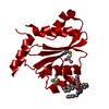
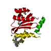
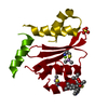
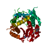
[English] 日本語
Yorodumi
- PDB-5oi3: Dissociation of biochemical and antiretroviral activities of Inte... -

+
Open data


ID or keywords:

Loading...

-
Basic information

Entry
Database: PDB / ID: 5oi3
TitleDissociation of biochemical and antiretroviral activities of Integrase-LEDGF Allosteric Inhibitors revealed by resistance of A125 polymorphic HIV-1
ComponentsIntegrase
KeywordsVIRAL PROTEIN / HIV-1 / Integrase / Catalytic core domain
Function / homology
Function and homology information


nucleotidyltransferase activity / : / : / HIV-1 retropepsin / retroviral ribonuclease H / exoribonuclease H / exoribonuclease H activity / host multivesicular body / DNA integration / RNA-directed DNA polymerase ...nucleotidyltransferase activity / : / : / HIV-1 retropepsin / retroviral ribonuclease H / exoribonuclease H / exoribonuclease H activity / host multivesicular body / DNA integration / RNA-directed DNA polymerase / viral genome integration into host DNA / viral penetration into host nucleus / establishment of integrated proviral latency / RNA-directed DNA polymerase activity / RNA-DNA hybrid ribonuclease activity / Transferases; Transferring phosphorus-containing groups; Nucleotidyltransferases / symbiont-mediated suppression of host gene expression / viral nucleocapsid / endonuclease activity / DNA recombination / nucleic acid binding / Hydrolases; Acting on ester bonds / DNA-directed DNA polymerase / aspartic-type endopeptidase activity / DNA-directed DNA polymerase activity / symbiont entry into host cell / lipid binding / host cell nucleus / host cell plasma membrane / virion membrane / structural molecule activity / proteolysis / DNA binding / RNA binding / zinc ion binding / membrane
Similarity search - Function
Ribonuclease H-like superfamily/Ribonuclease H / Reverse transcriptase connection / Reverse transcriptase connection domain / Reverse transcriptase thumb / Reverse transcriptase thumb domain / Integrase Zinc binding domain / Zinc finger integrase-type profile. / Integrase-like, N-terminal / Integrase DNA binding domain / Integrase, C-terminal domain superfamily, retroviral ...Ribonuclease H-like superfamily/Ribonuclease H / Reverse transcriptase connection / Reverse transcriptase connection domain / Reverse transcriptase thumb / Reverse transcriptase thumb domain / Integrase Zinc binding domain / Zinc finger integrase-type profile. / Integrase-like, N-terminal / Integrase DNA binding domain / Integrase, C-terminal domain superfamily, retroviral / Integrase, N-terminal zinc-binding domain / Integrase, C-terminal, retroviral / Integrase DNA binding domain profile. / Immunodeficiency lentiviral matrix, N-terminal / gag gene protein p17 (matrix protein) / RNase H / Integrase core domain / Integrase, catalytic core / Integrase catalytic domain profile. / Retroviral nucleocapsid Gag protein p24, C-terminal domain / Gag protein p24 C-terminal domain / Retropepsin-like catalytic domain / Matrix protein, lentiviral and alpha-retroviral, N-terminal / Ribonuclease H domain / RNase H type-1 domain profile. / Reverse transcriptase (RNA-dependent DNA polymerase) / Reverse transcriptase domain / Reverse transcriptase (RT) catalytic domain profile. / Retropepsins / Retroviral aspartyl protease / Aspartyl protease, retroviral-type family profile. / Peptidase A2A, retrovirus, catalytic / Retrovirus capsid, C-terminal / Retroviral matrix protein / Retrovirus capsid, N-terminal / zinc finger / Zinc knuckle / Zinc finger, CCHC-type superfamily / Zinc finger, CCHC-type / Zinc finger CCHC-type profile. / Nucleotidyltransferase; domain 5 / Aspartic peptidase, active site / Eukaryotic and viral aspartyl proteases active site. / Ribonuclease H superfamily / Aspartic peptidase domain superfamily / Ribonuclease H-like superfamily / Reverse transcriptase/Diguanylate cyclase domain / DNA/RNA polymerase superfamily / 2-Layer Sandwich / Alpha Beta
Similarity search - Domain/homology
Integrase / Gag-Pol polyprotein
Similarity search - Component
Biological speciesHuman immunodeficiency virus 1
MethodX-RAY DIFFRACTION / SYNCHROTRON / MOLECULAR REPLACEMENT / Resolution: 2.3 Å
AuthorsRuff, M. / Benarous, R.
CitationJournal: J. Biol. Chem. / Year: 2018
Title: Structure-function analyses unravel distinct effects of allosteric inhibitors of HIV-1 integrase on viral maturation and integration.
Authors: Bonnard, D. / Le Rouzic, E. / Eiler, S. / Amadori, C. / Orlov, I. / Bruneau, J.M. / Brias, J. / Barbion, J. / Chevreuil, F. / Spehner, D. / Chasset, S. / Ledoussal, B. / Moreau, F. / Saib, A. ...Authors: Bonnard, D. / Le Rouzic, E. / Eiler, S. / Amadori, C. / Orlov, I. / Bruneau, J.M. / Brias, J. / Barbion, J. / Chevreuil, F. / Spehner, D. / Chasset, S. / Ledoussal, B. / Moreau, F. / Saib, A. / Klaholz, B.P. / Emiliani, S. / Ruff, M. / Zamborlini, A. / Benarous, R.
History
DepositionJul 18, 2017Deposition site: PDBE / Processing site: PDBE
Revision 1.0Mar 7, 2018Provider: repository / Type: Initial release
Revision 1.1Mar 14, 2018Group: Database references / Category: citation / citation_author
Item: _citation.country / _citation.journal_abbrev ..._citation.country / _citation.journal_abbrev / _citation.journal_id_ASTM / _citation.journal_id_CSD / _citation.journal_id_ISSN / _citation.pdbx_database_id_DOI / _citation.pdbx_database_id_PubMed / _citation.title / _citation.year
Revision 1.2May 2, 2018Group: Data collection / Database references / Category: citation
Item: _citation.journal_volume / _citation.page_first / _citation.page_last
Revision 1.3Oct 16, 2019Group: Data collection / Category: reflns_shell
Revision 1.4Jan 17, 2024Group: Data collection / Database references / Refinement description
Category: chem_comp_atom / chem_comp_bond ...chem_comp_atom / chem_comp_bond / database_2 / pdbx_initial_refinement_model
Item: _database_2.pdbx_DOI / _database_2.pdbx_database_accession

-
Structure visualization

Structure viewerMolecule:
MolmilJmol/JSmol

Downloads & links

-
Assembly

Deposited unit
A: Integrase
hetero molecules


Theoretical massNumber of molelcules
Total (without water)20,1042
Polymers20,0801
Non-polymers241
Water1,820101
1
A: Integrase
hetero molecules

A: Integrase
hetero molecules


Theoretical massNumber of molelcules
Total (without water)40,2084
Polymers40,1592
Non-polymers492
Water362
TypeNameSymmetry operationNumber
identity operation1_555x,y,z1
crystal symmetry operation6_555-x,-x+y,-z+1/31
Buried area3090 Å2
ΔGint-34 kcal/mol
Surface area11870 Å2
MethodPISA
Unit cell
Length a, b, c (Å)72.363, 72.363, 65.819
Angle α, β, γ (deg.)90.00, 90.00, 120.00
Int Tables number152
Space group name H-MP3121
Components on special symmetry positions
IDModelComponents
11A-435-

HOH

-
Components

#1: Protein Integrase /


Mass: 20079.594 Da / Num. of mol.: 1 / Fragment: UNP Residues 50-212
Source method: isolated from a genetically manipulated source
Source: (gene. exp.) Human immunodeficiency virus 1 / Gene: pol / Production host: Escherichia coli (E. coli) / References: UniProt: B9VID1, UniProt: P03366*PLUS
#2: Chemical ChemComp-MG / MAGNESIUM ION


Mass: 24.305 Da / Num. of mol.: 1 / Source method: obtained synthetically / Formula: Mg
#3: Water ChemComp-HOH / water / Water


Mass: 18.015 Da / Num. of mol.: 101 / Source method: isolated from a natural source / Formula: H2O

-
Experimental details

-
Experiment

ExperimentMethod: X-RAY DIFFRACTION / Number of used crystals: 1

-
Sample preparation

CrystalDensity Matthews: 2.5 Å3/Da / Density % sol: 50.87 %
Crystal growTemperature: 297 K / Method: vapor diffusion, hanging drop / pH: 6.5 / Details: Ammonium sulfate, cacodylate

-
Data collection

DiffractionMean temperature: 100 K
Diffraction sourceSource: SYNCHROTRON / Site: SLS / Beamline: X06DA / Wavelength: 1 Å
DetectorType: DECTRIS PILATUS 2M / Detector: PIXEL / Date: Mar 26, 2013
RadiationProtocol: SINGLE WAVELENGTH / Monochromatic (M) / Laue (L): M / Scattering type: x-ray
Radiation wavelengthWavelength: 1 Å / Relative weight: 1
ReflectionResolution: 1.9→38.18 Å / Num. obs: 26873 / % possible obs: 88.7 % / Redundancy: 3.3 % / Net I/σ(I): 12.37

-
Processing

Software
NameVersionClassification
REFMAC5.8.0135refinement
XDSdata reduction
XDSdata scaling
MOLREPphasing
RefinementMethod to determine structure: MOLECULAR REPLACEMENT
Starting model: 4LH4
Resolution: 2.3→36.18 Å / Cor.coef. Fo:Fc: 0.947 / Cor.coef. Fo:Fc free: 0.871 / SU B: 5.773 / SU ML: 0.143 / Cross valid method: THROUGHOUT / ESU R: 0.235 / ESU R Free: 0.23 / Stereochemistry target values: MAXIMUM LIKELIHOOD / Details: HYDROGENS HAVE BEEN ADDED IN THE RIDING POSITIONS
RfactorNum. reflection% reflectionSelection details
Rfree0.27197 456 5 %RANDOM
Rwork0.19244 ---
obs0.19635 8663 99.29 %-
Solvent computationIon probe radii: 0.8 Å / Shrinkage radii: 0.8 Å / VDW probe radii: 1.2 Å / Solvent model: MASK
Displacement parametersBiso mean: 47.516 Å2
Baniso -1Baniso -2Baniso -3
1-0.01 Å20.01 Å20 Å2
2--0.01 Å2-0 Å2
3----0.04 Å2
Refinement stepCycle: 1 / Resolution: 2.3→36.18 Å
ProteinNucleic acidLigandSolventTotal
Num. atoms1023 0 1 101 1125
Refine LS restraints
Refine-IDTypeDev idealDev ideal targetNumber
X-RAY DIFFRACTIONr_bond_refined_d0.0170.0191042
X-RAY DIFFRACTIONr_bond_other_d0.0020.021035
X-RAY DIFFRACTIONr_angle_refined_deg1.6821.9371409
X-RAY DIFFRACTIONr_angle_other_deg1.01932369
X-RAY DIFFRACTIONr_dihedral_angle_1_deg5.8355128
X-RAY DIFFRACTIONr_dihedral_angle_2_deg33.84224.52442
X-RAY DIFFRACTIONr_dihedral_angle_3_deg19.92515180
X-RAY DIFFRACTIONr_dihedral_angle_4_deg18.814154
X-RAY DIFFRACTIONr_chiral_restr0.1070.2164
X-RAY DIFFRACTIONr_gen_planes_refined0.0080.021146
X-RAY DIFFRACTIONr_gen_planes_other0.0010.02232
X-RAY DIFFRACTIONr_nbd_refined
X-RAY DIFFRACTIONr_nbd_other
X-RAY DIFFRACTIONr_nbtor_refined
X-RAY DIFFRACTIONr_nbtor_other
X-RAY DIFFRACTIONr_xyhbond_nbd_refined
X-RAY DIFFRACTIONr_xyhbond_nbd_other
X-RAY DIFFRACTIONr_metal_ion_refined
X-RAY DIFFRACTIONr_metal_ion_other
X-RAY DIFFRACTIONr_symmetry_vdw_refined
X-RAY DIFFRACTIONr_symmetry_vdw_other
X-RAY DIFFRACTIONr_symmetry_hbond_refined
X-RAY DIFFRACTIONr_symmetry_hbond_other
X-RAY DIFFRACTIONr_symmetry_metal_ion_refined
X-RAY DIFFRACTIONr_symmetry_metal_ion_other
X-RAY DIFFRACTIONr_mcbond_it4.2144.222521
X-RAY DIFFRACTIONr_mcbond_other4.2124.216520
X-RAY DIFFRACTIONr_mcangle_it6.0876.283646
X-RAY DIFFRACTIONr_mcangle_other6.0846.289647
X-RAY DIFFRACTIONr_scbond_it5.3084.969521
X-RAY DIFFRACTIONr_scbond_other5.3034.972522
X-RAY DIFFRACTIONr_scangle_it
X-RAY DIFFRACTIONr_scangle_other8.2047.122764
X-RAY DIFFRACTIONr_long_range_B_refined13.69835.3941251
X-RAY DIFFRACTIONr_long_range_B_other13.72734.8351218
X-RAY DIFFRACTIONr_rigid_bond_restr
X-RAY DIFFRACTIONr_sphericity_free
X-RAY DIFFRACTIONr_sphericity_bonded
LS refinement shellResolution: 2.3→2.359 Å / Total num. of bins used: 20
RfactorNum. reflection% reflection
Rfree0.358 34 -
Rwork0.297 645 -
obs--100 %

+
About Yorodumi

-
News

-
Feb 9, 2022. New format data for meta-information of EMDB entries

New format data for meta-information of EMDB entries

  • Version 3 of the EMDB header file is now the official format.
  • The previous official version 1.9 will be removed from the archive.

Related info.:EMDB header

External links:wwPDB to switch to version 3 of the EMDB data model

-
Aug 12, 2020. Covid-19 info

Covid-19 info

URL: https://pdbj.org/emnavi/covid19.php

New page: Covid-19 featured information page in EM Navigator.

Related info.:Covid-19 info / Mar 5, 2020. Novel coronavirus structure data

+
Mar 5, 2020. Novel coronavirus structure data

Novel coronavirus structure data

Related info.:Yorodumi Speices / Aug 12, 2020. Covid-19 info

External links:COVID-19 featured content - PDBj / Molecule of the Month (242):Coronavirus Proteases

+
Jan 31, 2019. EMDB accession codes are about to change! (news from PDBe EMDB page)

EMDB accession codes are about to change! (news from PDBe EMDB page)

  • The allocation of 4 digits for EMDB accession codes will soon come to an end. Whilst these codes will remain in use, new EMDB accession codes will include an additional digit and will expand incrementally as the available range of codes is exhausted. The current 4-digit format prefixed with “EMD-” (i.e. EMD-XXXX) will advance to a 5-digit format (i.e. EMD-XXXXX), and so on. It is currently estimated that the 4-digit codes will be depleted around Spring 2019, at which point the 5-digit format will come into force.
  • The EM Navigator/Yorodumi systems omit the EMD- prefix.

Related info.:Q: What is EMD? / ID/Accession-code notation in Yorodumi/EM Navigator

External links:EMDB Accession Codes are Changing Soon! / Contact to PDBj

+
Jul 12, 2017. Major update of PDB

Major update of PDB

  • wwPDB released updated PDB data conforming to the new PDBx/mmCIF dictionary.
  • This is a major update changing the version number from 4 to 5, and with Remediation, in which all the entries are updated.
  • In this update, many items about electron microscopy experimental information are reorganized (e.g. em_software).
  • Now, EM Navigator and Yorodumi are based on the updated data.

External links:wwPDB Remediation / Enriched Model Files Conforming to OneDep Data Standards Now Available in the PDB FTP Archive

-
Yorodumi

Thousand views of thousand structures

  • Yorodumi is a browser for structure data from EMDB, PDB, SASBDB, etc.
  • This page is also the successor to EM Navigator detail page, and also detail information page/front-end page for Omokage search.
  • The word "yorodu" (or yorozu) is an old Japanese word meaning "ten thousand". "mi" (miru) is to see.

Related info.:EMDB / PDB / SASBDB / Comparison of 3 databanks / Yorodumi Search / Aug 31, 2016. New EM Navigator & Yorodumi / Yorodumi Papers / Jmol/JSmol / Function and homology information / Changes in new EM Navigator and Yorodumi

Read more