 Movie
Movie Controller


+ Open data

- Basic information
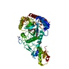
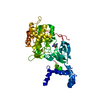
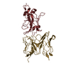
| Entry | Database: PDB / ID: 5o9p | ||||||
|---|---|---|---|---|---|---|---|
| Title | Crystal structure of Gas2 in complex with compound 10 | ||||||
 Components Components | 1,3-beta-glucanosyltransferase GAS2 | ||||||
 Keywords Keywords |  TRANSFERASE / Aspergillus fumigatus / AfGel4 / ScGas2 / transglycosylases / glucanosyltransferases / cell wall remodeling / fungal cell wall TRANSFERASE / Aspergillus fumigatus / AfGel4 / ScGas2 / transglycosylases / glucanosyltransferases / cell wall remodeling / fungal cell wall | ||||||
| Function / homology |  Function and homology information Function and homology information1,3-beta-glucanosyltransferase activity / fungal-type cell wall (1->3)-beta-D-glucan biosynthetic process / ascospore wall assembly / fungal-type cell wall organization / fungal-type vacuole / Transferases; Glycosyltransferases; Hexosyltransferases / side of membrane / plasma membrane / cytoplasmSimilarity search - Function | ||||||
| Biological species |  Saccharomyces cerevisiae S288c (yeast) Saccharomyces cerevisiae S288c (yeast) | ||||||
| Method | X-RAY DIFFRACTION / SYNCHROTRON / MOLECULAR REPLACEMENT / Resolution: 1.75 Å | ||||||
 Authors Authors | Delso, I. / Valero-Gonzalez, J. / Gomollon-Bel, F. / Castro-Lopez, J. / Fang, W. / Navratilova, I. / Van Aalten, D. / Tejero, T. / Merino, P. / Hurtado-Guerrero, R. | ||||||
 Citation Citation | Journal: ChemMedChem / Year: 2018 Title: Inhibitors against Fungal Cell Wall Remodeling Enzymes. Authors: Delso, I. / Valero-Gonzalez, J. / Gomollon-Bel, F. / Castro-Lopez, J. / Fang, W. / Navratilova, I. / van Aalten, D.M.F. / Tejero, T. / Merino, P. / Hurtado-Guerrero, R. | ||||||
| History |
|
- Structure visualization
| Structure viewer | Molecule: MolmilJmol/JSmol |
|---|
- Downloads & links
-Download
| PDBx/mmCIF format | 5o9p.cif.gz | 199.4 KB | Display | PDBx/mmCIF format |
|---|---|---|---|---|
| PDB format | pdb5o9p.ent.gz | 157.6 KB | Display | PDB format |
| PDBx/mmJSON format | 5o9p.json.gz | Tree view | PDBx/mmJSON format | |
| Others |  Other downloads Other downloads |
-Validation report
| Arichive directory | https://data.pdbj.org/pub/pdb/validation_reports/o9/5o9pftp://data.pdbj.org/pub/pdb/validation_reports/o9/5o9p | HTTPS FTP |
|---|
-Related structure data
| Related structure data |  5o9oC  5o9qC  5o9rC  5o9yC  5oa2C  5oa6C 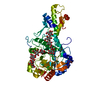 2w62S S: Starting model for refinement C: citing same article ( |
|---|---|
| Similar structure data |


-Links
 PDBj
PDBj- Assembly
| Deposited unit | 
| ||||||||
|---|---|---|---|---|---|---|---|---|---|
| 1 |
| ||||||||
| Unit cell |
|
-Components
| #1: Protein | Mass: 62426.000 Da / Num. of mol.: 1 Source method: isolated from a genetically manipulated source Source: (gene. exp.) Saccharomyces cerevisiae S288c (yeast) / Gene: GAS2, YLR343W, L8300.5 / Production host:  Komagataella pastoris (fungus) Komagataella pastoris (fungus)References: UniProt: Q06135, Transferases; Glycosyltransferases; Hexosyltransferases |
|---|---|
| #2: Chemical | ChemComp-9P8 / (  Mass: 676.624 Da / Num. of mol.: 1 / Source method: obtained synthetically / Formula: C27H40N4O16 Mass: 676.624 Da / Num. of mol.: 1 / Source method: obtained synthetically / Formula: C27H40N4O16 |
| #3: Chemical | ChemComp-EDO / Ethylene glycol  Mass: 62.068 Da / Num. of mol.: 1 / Source method: obtained synthetically / Formula: C2H6O2 Mass: 62.068 Da / Num. of mol.: 1 / Source method: obtained synthetically / Formula: C2H6O2 |
| #4: Water | ChemComp-HOH / Water Mass: 18.015 Da / Num. of mol.: 262 / Source method: isolated from a natural source / Formula: H2O Mass: 18.015 Da / Num. of mol.: 262 / Source method: isolated from a natural source / Formula: H2O |
-Experimental details
-Experiment
| Experiment | Method: X-RAY DIFFRACTION / Number of used crystals: 1 |
|---|
- Sample preparation
| Crystal | Density Matthews: 2.15 Å3/Da / Density % sol: 42.74 % |
|---|---|
| Crystal grow | Temperature: 291 K / Method: vapor diffusion, sitting drop / Details: PEG 3500, ammonium sulfate, BIS-Tris pH 6 |
-Data collection
| Diffraction | Mean temperature: 100 K |
|---|---|
| Diffraction source | Source: SYNCHROTRON / Site: ALBA  / Beamline: XALOC / Wavelength: 0.979 Å / Beamline: XALOC / Wavelength: 0.979 Å |
| Detector | Type: DECTRIS PILATUS3 S 6M / Detector: PIXEL / Date: Oct 10, 2014 |
| Radiation | Protocol: SINGLE WAVELENGTH / Monochromatic (M) / Laue (L): M / Scattering type: x-ray |
| Radiation wavelength | Wavelength: 0.979 Å / Relative weight: 1 |
| Reflection | Resolution: 1.75→75.5 Å / Num. obs: 54873 / % possible obs: 99.6 % / Redundancy: 5.9 % / Rmerge(I) obs: 0.164 / Net I/σ(I): 5.7 |
| Reflection shell | Resolution: 1.75→1.85 Å / Redundancy: 6.3 % / Rmerge(I) obs: 0.555 / Mean I/σ(I) obs: 2 / % possible all: 100 |
- Processing
| Software |
| ||||||||||||||||||||||||||||||||||||||||||||||||||||||||||||||||||||||||||||||||||||||||||||||||||||||||||||||||||||||||||||||||||||||||||||||||||||||||||||||||||||||||||||||||||||||
|---|---|---|---|---|---|---|---|---|---|---|---|---|---|---|---|---|---|---|---|---|---|---|---|---|---|---|---|---|---|---|---|---|---|---|---|---|---|---|---|---|---|---|---|---|---|---|---|---|---|---|---|---|---|---|---|---|---|---|---|---|---|---|---|---|---|---|---|---|---|---|---|---|---|---|---|---|---|---|---|---|---|---|---|---|---|---|---|---|---|---|---|---|---|---|---|---|---|---|---|---|---|---|---|---|---|---|---|---|---|---|---|---|---|---|---|---|---|---|---|---|---|---|---|---|---|---|---|---|---|---|---|---|---|---|---|---|---|---|---|---|---|---|---|---|---|---|---|---|---|---|---|---|---|---|---|---|---|---|---|---|---|---|---|---|---|---|---|---|---|---|---|---|---|---|---|---|---|---|---|---|---|---|---|
| Refinement | Method to determine structure: MOLECULAR REPLACEMENT Starting model: 2W62 Resolution: 1.75→75.5 Å / Cor.coef. Fo:Fc: 0.937 / Cor.coef. Fo:Fc free: 0.919 / SU B: 6.675 / SU ML: 0.095 / Cross valid method: THROUGHOUT / ESU R: 0.119 / ESU R Free: 0.116 / Details: HYDROGENS HAVE BEEN ADDED IN THE RIDING POSITIONS
| ||||||||||||||||||||||||||||||||||||||||||||||||||||||||||||||||||||||||||||||||||||||||||||||||||||||||||||||||||||||||||||||||||||||||||||||||||||||||||||||||||||||||||||||||||||||
| Solvent computation | Ion probe radii: 0.8 Å / Shrinkage radii: 0.8 Å / VDW probe radii: 1.2 Å | ||||||||||||||||||||||||||||||||||||||||||||||||||||||||||||||||||||||||||||||||||||||||||||||||||||||||||||||||||||||||||||||||||||||||||||||||||||||||||||||||||||||||||||||||||||||
| Displacement parameters | Biso mean: 26.772 Å2
| ||||||||||||||||||||||||||||||||||||||||||||||||||||||||||||||||||||||||||||||||||||||||||||||||||||||||||||||||||||||||||||||||||||||||||||||||||||||||||||||||||||||||||||||||||||||
| Refinement step | Cycle: 1 / Resolution: 1.75→75.5 Å
| ||||||||||||||||||||||||||||||||||||||||||||||||||||||||||||||||||||||||||||||||||||||||||||||||||||||||||||||||||||||||||||||||||||||||||||||||||||||||||||||||||||||||||||||||||||||
| Refine LS restraints |
|