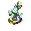
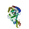
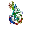
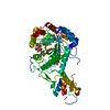
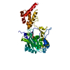
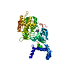
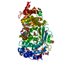
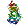
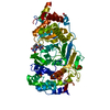
[English] 日本語
Yorodumi
- PDB-2w62: Saccharomyces cerevisiae Gas2p in complex with laminaripentaose -

+
Open data


ID or keywords:

Loading...

-
Basic information

Entry
Database: PDB / ID: 2w62
TitleSaccharomyces cerevisiae Gas2p in complex with laminaripentaose
ComponentsGLYCOLIPID-ANCHORED SURFACE PROTEIN 2
KeywordsTRANSFERASE / GLYCOPROTEIN / CELL MEMBRANE / FUNGAL CELL WALL / TRANSGLYCOSYLATION / GLUCAN / MEMBRANE / GPI-ANCHOR / LIPOPROTEIN
Function / homology
Function and homology information


1,3-beta-glucanosyltransferase activity / fungal-type cell wall (1->3)-beta-D-glucan biosynthetic process / ascospore wall assembly / fungal-type cell wall organization / fungal-type vacuole / Transferases; Glycosyltransferases; Hexosyltransferases / side of membrane / plasma membrane / cytoplasm
Similarity search - Function
Methane Monooxygenase Hydroxylase; Chain G, domain 1 - #1040 / Glucanosyltransferase / Glucanosyltransferase / X8 domain / X8 domain / Possibly involved in carbohydrate binding / Methane Monooxygenase Hydroxylase; Chain G, domain 1 / Glycosidases / Glycoside hydrolase superfamily / TIM Barrel ...Methane Monooxygenase Hydroxylase; Chain G, domain 1 - #1040 / Glucanosyltransferase / Glucanosyltransferase / X8 domain / X8 domain / Possibly involved in carbohydrate binding / Methane Monooxygenase Hydroxylase; Chain G, domain 1 / Glycosidases / Glycoside hydrolase superfamily / TIM Barrel / Alpha-Beta Barrel / Up-down Bundle / Mainly Alpha / Alpha Beta
Similarity search - Domain/homology
1,4-BUTANEDIOL / 1,3-beta-glucanosyltransferase GAS2
Similarity search - Component
Biological speciesSACCHAROMYCES CEREVISIAE (brewer's yeast)
MethodX-RAY DIFFRACTION / MOLECULAR REPLACEMENT / Resolution: 1.85 Å
AuthorsSchuettelkopf, A.W. / Hurtado-Guerrero, R. / van Aalten, D.M.F.
CitationJournal: J.Biol.Chem. / Year: 2009
Title: Molecular Mechanisms of Yeast Cell Wall Glucan Remodeling.
Authors: Hurtado-Guerrero, R. / Schuttelkopf, A.W. / Mouyna, I. / Ibrahim, A.F.M. / Shepherd, S. / Fontaine, T. / Latge, J. / Van Aalten, D.M.F.
History
DepositionDec 16, 2008Deposition site: PDBE / Processing site: PDBE
Revision 1.0Jan 27, 2009Provider: repository / Type: Initial release
Revision 1.1Jul 13, 2011Group: Advisory / Version format compliance
Revision 1.2Aug 17, 2011Group: Atomic model / Derived calculations ...Atomic model / Derived calculations / Non-polymer description / Refinement description / Structure summary
Revision 1.3Feb 8, 2012Group: Other
Revision 2.0Jul 29, 2020Group: Atomic model / Data collection ...Atomic model / Data collection / Derived calculations / Other / Structure summary
Category: atom_site / chem_comp ...atom_site / chem_comp / entity / pdbx_branch_scheme / pdbx_chem_comp_identifier / pdbx_database_status / pdbx_entity_branch / pdbx_entity_branch_descriptor / pdbx_entity_branch_link / pdbx_entity_branch_list / pdbx_entity_nonpoly / pdbx_nonpoly_scheme / pdbx_struct_assembly_gen / struct_asym / struct_conn / struct_site / struct_site_gen
Item: _atom_site.auth_asym_id / _atom_site.auth_seq_id ..._atom_site.auth_asym_id / _atom_site.auth_seq_id / _atom_site.label_asym_id / _chem_comp.name / _chem_comp.type / _entity.formula_weight / _entity.pdbx_description / _entity.pdbx_number_of_molecules / _entity.type / _pdbx_database_status.status_code_sf / _pdbx_struct_assembly_gen.asym_id_list / _struct_conn.pdbx_leaving_atom_flag / _struct_conn.ptnr1_auth_asym_id / _struct_conn.ptnr1_auth_seq_id / _struct_conn.ptnr1_label_asym_id / _struct_conn.ptnr2_auth_asym_id / _struct_conn.ptnr2_auth_seq_id / _struct_conn.ptnr2_label_asym_id
Description: Carbohydrate remediation / Provider: repository / Type: Remediation
Revision 2.1May 1, 2024Group: Data collection / Database references ...Data collection / Database references / Refinement description / Structure summary
Category: chem_comp / chem_comp_atom ...chem_comp / chem_comp_atom / chem_comp_bond / database_2 / pdbx_initial_refinement_model
Item: _chem_comp.pdbx_synonyms / _database_2.pdbx_DOI / _database_2.pdbx_database_accession
Remark 700 SHEET DETERMINATION METHOD: DSSP THE SHEETS PRESENTED AS "AB" IN EACH CHAIN ON SHEET RECORDS BELOW ... SHEET DETERMINATION METHOD: DSSP THE SHEETS PRESENTED AS "AB" IN EACH CHAIN ON SHEET RECORDS BELOW IS ACTUALLY AN 8-STRANDED BARREL THIS IS REPRESENTED BY A 9-STRANDED SHEET IN WHICH THE FIRST AND LAST STRANDS ARE IDENTICAL.

-
Structure visualization

Structure viewerMolecule:
MolmilJmol/JSmol

Downloads & links

-
Assembly

Deposited unit
A: GLYCOLIPID-ANCHORED SURFACE PROTEIN 2
hetero molecules


Theoretical massNumber of molelcules
Total (without water)64,1744
Polymers62,4261
Non-polymers1,7483
Water5,639313
1


  • Idetical with deposited unit
  • defined by author&software
TypeNameSymmetry operationNumber
identity operation1_555x,y,z1
MethodPISA
Unit cell
Length a, b, c (Å)50.039, 70.841, 149.149
Angle α, β, γ (deg.)90.00, 90.00, 90.00
Int Tables number19
Space group name H-MP212121

-
Components

#1: Protein GLYCOLIPID-ANCHORED SURFACE PROTEIN 2 / GAS2P


Mass: 62426.000 Da / Num. of mol.: 1
Source method: isolated from a genetically manipulated source
Source: (gene. exp.) SACCHAROMYCES CEREVISIAE (brewer's yeast)
Strain: W303 / Plasmid: PPICZALPHA-BASED / Production host: PICHIA PASTORIS (fungus) / Strain (production host): X33
References: UniProt: Q06135, Transferases; Glycosyltransferases; Hexosyltransferases
#2: Polysaccharide beta-D-glucopyranose-(1-3)-beta-D-glucopyranose-(1-3)-beta-D-glucopyranose-(1-3)-beta-D- ...beta-D-glucopyranose-(1-3)-beta-D-glucopyranose-(1-3)-beta-D-glucopyranose-(1-3)-beta-D-glucopyranose-(1-3)-beta-D-glucopyranose


Type: oligosaccharide / Mass: 828.719 Da / Num. of mol.: 2
Source method: isolated from a genetically manipulated source
DescriptorTypeProgram
DGlcpb1-3DGlcpb1-3DGlcpb1-3DGlcpb1-3DGlcpb1-ROHGlycam Condensed SequenceGMML 1.0
WURCS=2.0/1,5,4/[a2122h-1b_1-5]/1-1-1-1-1/a3-b1_b3-c1_c3-d1_d3-e1WURCSPDB2Glycan 1.1.0
[][b-D-Glcp]{[(3+1)][b-D-Glcp]{[(3+1)][b-D-Glcp]{[(3+1)][b-D-Glcp]{[(3+1)][b-D-Glcp]{}}}}}LINUCSPDB-CARE
#3: Chemical ChemComp-BU1 / 1,4-BUTANEDIOL / 1,4-Butanediol


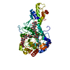
Mass: 90.121 Da / Num. of mol.: 1 / Source method: obtained synthetically / Formula: C4H10O2
#4: Water ChemComp-HOH / water / Water


Mass: 18.015 Da / Num. of mol.: 313 / Source method: isolated from a natural source / Formula: H2O
Sequence detailsMISSING RESIDUES WERE NOT MODELLED DUE TO LACKING ELECTRON DENSITY

-
Experimental details

-
Experiment

ExperimentMethod: X-RAY DIFFRACTION / Number of used crystals: 1

-
Sample preparation

CrystalDensity Matthews: 2 Å3/Da / Density % sol: 40 % / Description: NONE
Crystal growDetails: 100 MM SODIUM ACETATE PH 4.5; 5% ACETONE; 20% 1,4-BUTANEDIOL

-
Data collection

DiffractionMean temperature: 100 K
Diffraction sourceSource: ROTATING ANODE / Type: RIGAKU RU200 / Wavelength: 1.5418
DetectorType: RIGAKU
RadiationProtocol: SINGLE WAVELENGTH / Monochromatic (M) / Laue (L): M / Scattering type: x-ray
Radiation wavelengthWavelength: 1.5418 Å / Relative weight: 1
ReflectionResolution: 1.85→20 Å / Num. obs: 45921 / % possible obs: 99.7 % / Observed criterion σ(I): 0 / Redundancy: 5.3 % / Rmerge(I) obs: 0.04 / Net I/σ(I): 23.3
Reflection shellResolution: 1.85→1.92 Å / Redundancy: 5.2 % / Rmerge(I) obs: 0.29 / Mean I/σ(I) obs: 5.5 / % possible all: 100

-
Processing

Software
NameVersionClassification
REFMAC5.4.0069refinement
DENZOdata reduction
SCALEPACKdata scaling
RefinementMethod to determine structure: MOLECULAR REPLACEMENT
Starting model: DERIVATIVE MODEL, NOT DEPOSITED

Resolution: 1.85→23 Å / Cor.coef. Fo:Fc: 0.959 / Cor.coef. Fo:Fc free: 0.952 / SU B: 5.048 / SU ML: 0.08 / TLS residual ADP flag: LIKELY RESIDUAL / Cross valid method: THROUGHOUT / ESU R: 0.124 / ESU R Free: 0.115 / Stereochemistry target values: MAXIMUM LIKELIHOOD / Details: HYDROGENS HAVE BEEN ADDED IN THE RIDING POSITIONS.
RfactorNum. reflection% reflectionSelection details
Rfree0.21 555 1.2 %RANDOM
Rwork0.183 ---
obs0.184 45304 99.5 %-
Solvent computationIon probe radii: 0.8 Å / Shrinkage radii: 0.8 Å / VDW probe radii: 1.2 Å / Solvent model: BABINET MODEL WITH MASK
Displacement parametersBiso mean: 42.47 Å2
Baniso -1Baniso -2Baniso -3
1--1.26 Å20 Å20 Å2
2--0.21 Å20 Å2
3---1.05 Å2
Refinement stepCycle: LAST / Resolution: 1.85→23 Å
ProteinNucleic acidLigandSolventTotal
Num. atoms3474 0 118 313 3905
Refine LS restraints
Refine-IDTypeDev idealDev ideal targetNumber
X-RAY DIFFRACTIONr_bond_refined_d0.0120.0223721
X-RAY DIFFRACTIONr_bond_other_d
X-RAY DIFFRACTIONr_angle_refined_deg1.3092.0335059
X-RAY DIFFRACTIONr_angle_other_deg
X-RAY DIFFRACTIONr_dihedral_angle_1_deg9.8785443
X-RAY DIFFRACTIONr_dihedral_angle_2_deg32.92724.746177
X-RAY DIFFRACTIONr_dihedral_angle_3_deg13.92515602
X-RAY DIFFRACTIONr_dihedral_angle_4_deg15.2421515
X-RAY DIFFRACTIONr_chiral_restr0.0930.2564
X-RAY DIFFRACTIONr_gen_planes_refined0.0050.0212779
X-RAY DIFFRACTIONr_gen_planes_other
X-RAY DIFFRACTIONr_nbd_refined
X-RAY DIFFRACTIONr_nbd_other
X-RAY DIFFRACTIONr_nbtor_refined
X-RAY DIFFRACTIONr_nbtor_other
X-RAY DIFFRACTIONr_xyhbond_nbd_refined
X-RAY DIFFRACTIONr_xyhbond_nbd_other
X-RAY DIFFRACTIONr_metal_ion_refined
X-RAY DIFFRACTIONr_metal_ion_other
X-RAY DIFFRACTIONr_symmetry_vdw_refined
X-RAY DIFFRACTIONr_symmetry_vdw_other
X-RAY DIFFRACTIONr_symmetry_hbond_refined
X-RAY DIFFRACTIONr_symmetry_hbond_other
X-RAY DIFFRACTIONr_symmetry_metal_ion_refined
X-RAY DIFFRACTIONr_symmetry_metal_ion_other
X-RAY DIFFRACTIONr_mcbond_it0.8391.52187
X-RAY DIFFRACTIONr_mcbond_other
X-RAY DIFFRACTIONr_mcangle_it1.46823534
X-RAY DIFFRACTIONr_mcangle_other
X-RAY DIFFRACTIONr_scbond_it2.26931534
X-RAY DIFFRACTIONr_scbond_other
X-RAY DIFFRACTIONr_scangle_it3.4414.51519
X-RAY DIFFRACTIONr_scangle_other
X-RAY DIFFRACTIONr_long_range_B_refined
X-RAY DIFFRACTIONr_long_range_B_other
X-RAY DIFFRACTIONr_rigid_bond_restr
X-RAY DIFFRACTIONr_sphericity_free
X-RAY DIFFRACTIONr_sphericity_bonded
LS refinement shellResolution: 1.85→1.9 Å / Total num. of bins used: 20 /
RfactorNum. reflection
Rfree0.281 48
Rwork0.201 3182
Refinement TLS params.Method: refined / Origin x: 5.4387 Å / Origin y: 27.3502 Å / Origin z: 19.5125 Å
111213212223313233
T-0.1586 Å20.048 Å20.0155 Å2--0.1446 Å2-0.0122 Å2---0.2062 Å2
L1.9553 °20.3959 °20.4071 °2-1.3753 °20.5677 °2--1.2669 °2
S0.0147 Å °-0.0961 Å °0.1689 Å °0.0367 Å °0.0083 Å °-0.0971 Å °0.119 Å °0.0141 Å °-0.023 Å °
Refinement TLS group
IDRefine-IDRefine TLS-IDAuth asym-IDAuth seq-ID
1X-RAY DIFFRACTION1A29 - 64
2X-RAY DIFFRACTION1A75 - 355
3X-RAY DIFFRACTION1A363 - 404
4X-RAY DIFFRACTION1A409 - 482
5X-RAY DIFFRACTION1A489
6X-RAY DIFFRACTION1A1490 - 1491

+
About Yorodumi

-
News

-
Feb 9, 2022. New format data for meta-information of EMDB entries

New format data for meta-information of EMDB entries

  • Version 3 of the EMDB header file is now the official format.
  • The previous official version 1.9 will be removed from the archive.

Related info.:EMDB header

External links:wwPDB to switch to version 3 of the EMDB data model

-
Aug 12, 2020. Covid-19 info

Covid-19 info

URL: https://pdbj.org/emnavi/covid19.php

New page: Covid-19 featured information page in EM Navigator.

Related info.:Covid-19 info / Mar 5, 2020. Novel coronavirus structure data

+
Mar 5, 2020. Novel coronavirus structure data

Novel coronavirus structure data

Related info.:Yorodumi Speices / Aug 12, 2020. Covid-19 info

External links:COVID-19 featured content - PDBj / Molecule of the Month (242):Coronavirus Proteases

+
Jan 31, 2019. EMDB accession codes are about to change! (news from PDBe EMDB page)

EMDB accession codes are about to change! (news from PDBe EMDB page)

  • The allocation of 4 digits for EMDB accession codes will soon come to an end. Whilst these codes will remain in use, new EMDB accession codes will include an additional digit and will expand incrementally as the available range of codes is exhausted. The current 4-digit format prefixed with “EMD-” (i.e. EMD-XXXX) will advance to a 5-digit format (i.e. EMD-XXXXX), and so on. It is currently estimated that the 4-digit codes will be depleted around Spring 2019, at which point the 5-digit format will come into force.
  • The EM Navigator/Yorodumi systems omit the EMD- prefix.

Related info.:Q: What is EMD? / ID/Accession-code notation in Yorodumi/EM Navigator

External links:EMDB Accession Codes are Changing Soon! / Contact to PDBj

+
Jul 12, 2017. Major update of PDB

Major update of PDB

  • wwPDB released updated PDB data conforming to the new PDBx/mmCIF dictionary.
  • This is a major update changing the version number from 4 to 5, and with Remediation, in which all the entries are updated.
  • In this update, many items about electron microscopy experimental information are reorganized (e.g. em_software).
  • Now, EM Navigator and Yorodumi are based on the updated data.

External links:wwPDB Remediation / Enriched Model Files Conforming to OneDep Data Standards Now Available in the PDB FTP Archive

-
Yorodumi

Thousand views of thousand structures

  • Yorodumi is a browser for structure data from EMDB, PDB, SASBDB, etc.
  • This page is also the successor to EM Navigator detail page, and also detail information page/front-end page for Omokage search.
  • The word "yorodu" (or yorozu) is an old Japanese word meaning "ten thousand". "mi" (miru) is to see.

Related info.:EMDB / PDB / SASBDB / Comparison of 3 databanks / Yorodumi Search / Aug 31, 2016. New EM Navigator & Yorodumi / Yorodumi Papers / Jmol/JSmol / Function and homology information / Changes in new EM Navigator and Yorodumi

Read more