 Movie
Movie Controller

[English] 日本語
 Yorodumi
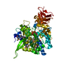
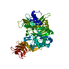
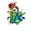
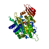
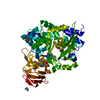
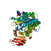
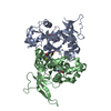
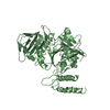
- PDB-5nzk: Crystal structure of UDP-glucose pyrophosphorylase from Leishmani... -
+ Open data

- Basic information
| Entry | Database: PDB / ID: 5nzk | ||||||
|---|---|---|---|---|---|---|---|
| Title | Crystal structure of UDP-glucose pyrophosphorylase from Leishmania major in complex with phenylalanine | ||||||
 Components Components | UDP-glucose pyrophosphorylase UTP—glucose-1-phosphate uridylyltransferase UTP—glucose-1-phosphate uridylyltransferase | ||||||
 Keywords Keywords | TRANSFERASE / NTP-transferase / Pathogen / Allostery / Catalysis | ||||||
| Function / homology |  Function and homology informationUTP-glucose-1-phosphate uridylyltransferase / UTP:glucose-1-phosphate uridylyltransferase activity / UDP-glucose metabolic process / nuclear lumen / ciliary plasm / glycogen metabolic process / cytosol / cytoplasm Function and homology informationUTP-glucose-1-phosphate uridylyltransferase / UTP:glucose-1-phosphate uridylyltransferase activity / UDP-glucose metabolic process / nuclear lumen / ciliary plasm / glycogen metabolic process / cytosol / cytoplasmSimilarity search - Function | ||||||
| Biological species |  Leishmania major (eukaryote) Leishmania major (eukaryote) | ||||||
| Method | X-RAY DIFFRACTION / SYNCHROTRON / MOLECULAR REPLACEMENT / Resolution: 2.45 Å | ||||||
 Authors Authors | Cramer, J.T. / Fuehring, J.I. / Baruch, P. / Bruetting, C. / Hesse, R. / Knoelker, H.-J. / Gerardy-Schahn, R. / Fedorov, R. | ||||||
| Funding support |  Germany, 1items Germany, 1items
| ||||||
 Citation Citation | Journal: Acs Catalysis / Year: 2018 Title: Decoding Allosteric Networks in Biocatalysts: Rational Approach to Therapies and Biotechnologies Authors: Cramer, J.T. / Fuehring, J.I. / Baruch, P. / Bruetting, C. / Knoelker, H.-J. / Gerardy-Schahn, R. / Fedorov, R. | ||||||
| History |
|
- Structure visualization
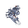
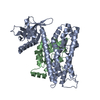
| Structure viewer | Molecule: MolmilJmol/JSmol |
|---|
- Downloads & links
-Download
| PDBx/mmCIF format | 5nzk.cif.gz | 109.7 KB | Display | PDBx/mmCIF format |
|---|---|---|---|---|
| PDB format | pdb5nzk.ent.gz | 83.3 KB | Display | PDB format |
| PDBx/mmJSON format | 5nzk.json.gz | Tree view | PDBx/mmJSON format | |
| Others |  Other downloads Other downloads |
-Validation report
| Arichive directory | https://data.pdbj.org/pub/pdb/validation_reports/nz/5nzkftp://data.pdbj.org/pub/pdb/validation_reports/nz/5nzk | HTTPS FTP |
|---|
-Related structure data
| Related structure data | 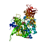 5nzgC 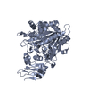 5nzhC 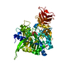 5nziC 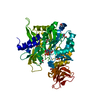 5nzjC  5nzlC 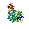 5nzmC  2oefS S: Starting model for refinement C: citing same article ( |
|---|---|
| Similar structure data |


-Links
 PDBj
PDBj


- Assembly
| Deposited unit | 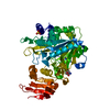
| ||||||||
|---|---|---|---|---|---|---|---|---|---|
| 1 |
| ||||||||
| Unit cell |
| ||||||||
| Components on special symmetry positions |
|

-Components
| #1: Protein | UTP—glucose-1-phosphate uridylyltransferase Mass: 56054.770 Da / Num. of mol.: 1 Source method: isolated from a genetically manipulated source Source: (gene. exp.) Leishmania major (eukaryote) / Gene: UGP, LMJF_18_0990 / Production host:  Escherichia coli (E. coli) Escherichia coli (E. coli)References: UniProt: Q4QDU3, UTP-glucose-1-phosphate uridylyltransferase |
|---|---|
| #2: Chemical | ChemComp-PHE / Phenylalanine  Type: L-peptide linking / Mass: 165.189 Da / Num. of mol.: 1 / Source method: obtained synthetically / Formula: C9H11NO2 Type: L-peptide linking / Mass: 165.189 Da / Num. of mol.: 1 / Source method: obtained synthetically / Formula: C9H11NO2 |
| #3: Chemical | ChemComp-SO4 / Sulfate  Mass: 96.063 Da / Num. of mol.: 1 / Source method: obtained synthetically / Formula: SO4 Mass: 96.063 Da / Num. of mol.: 1 / Source method: obtained synthetically / Formula: SO4 |
| #4: Water | ChemComp-HOH / Water Mass: 18.015 Da / Num. of mol.: 116 / Source method: isolated from a natural source / Formula: H2O Mass: 18.015 Da / Num. of mol.: 116 / Source method: isolated from a natural source / Formula: H2O |
-Experimental details
-Experiment
| Experiment | Method: X-RAY DIFFRACTION / Number of used crystals: 1 |
|---|
- Sample preparation
| Crystal | Density Matthews: 2.63 Å3/Da / Density % sol: 53.24 % / Mosaicity: 0.18 ° |
|---|---|
| Crystal grow | Temperature: 291.15 K / Method: vapor diffusion, sitting drop Details: 24% PEG 3350, 100 mM Bis-Tris pH 6.8, 0.2 M Li2SO4, 2 mM DTT |
-Data collection
| Diffraction | Mean temperature: 100 K |
|---|---|
| Diffraction source | Source: SYNCHROTRON / Site: ESRF  / Beamline: ID29 / Wavelength: 0.92 / Wavelength: 0.92 Å / Beamline: ID29 / Wavelength: 0.92 / Wavelength: 0.92 Å |
| Detector | Type: DECTRIS PILATUS 6M / Detector: PIXEL / Date: Nov 29, 2012 |
| Radiation | Protocol: SINGLE WAVELENGTH / Monochromatic (M) / Laue (L): M / Scattering type: x-ray |
| Radiation wavelength | Wavelength: 0.92 Å / Relative weight: 1 |
| Reflection | Resolution: 2.45→50 Å / Num. obs: 22170 / % possible obs: 100 % / Redundancy: 12.99 % / Biso Wilson estimate: 52 Å2 / Rsym value: 0.037 / Net I/σ(I): 17.27 |
| Reflection shell | Mean I/σ(I) obs: 2.28 / Num. unique all: 2219 / Rsym value: 0.4357 / % possible all: 100 |
- Processing
| Software |
| |||||||||||||||||||||||||||||||||||||||||||||||||||||||||||||||
|---|---|---|---|---|---|---|---|---|---|---|---|---|---|---|---|---|---|---|---|---|---|---|---|---|---|---|---|---|---|---|---|---|---|---|---|---|---|---|---|---|---|---|---|---|---|---|---|---|---|---|---|---|---|---|---|---|---|---|---|---|---|---|---|---|
| Refinement | Method to determine structure: MOLECULAR REPLACEMENT Starting model: 2OEF Resolution: 2.45→47.081 Å / SU ML: 0.3 / Cross valid method: FREE R-VALUE / σ(F): 1.34 / Phase error: 25.75
| |||||||||||||||||||||||||||||||||||||||||||||||||||||||||||||||
| Solvent computation | Shrinkage radii: 0.9 Å / VDW probe radii: 1.11 Å | |||||||||||||||||||||||||||||||||||||||||||||||||||||||||||||||
| Refinement step | Cycle: LAST / Resolution: 2.45→47.081 Å
| |||||||||||||||||||||||||||||||||||||||||||||||||||||||||||||||
| Refine LS restraints |
| |||||||||||||||||||||||||||||||||||||||||||||||||||||||||||||||
| LS refinement shell |
|
