 Movie
Movie Controller

[English] 日本語
 Yorodumi
- PDB-5nyk: Crystal structure of the atypical poplar thioredoxin-like2.1 in o... -
+ Open data

- Basic information
| Entry | Database: PDB / ID: 5nyk | |||||||||
|---|---|---|---|---|---|---|---|---|---|---|
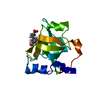
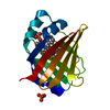
| Title | Crystal structure of the atypical poplar thioredoxin-like2.1 in oxidized state | |||||||||
 Components Components | Thioredoxin-like protein 2.1 | |||||||||
 Keywords Keywords |  OXIDOREDUCTASE / atypical thioredoxin / disulfide exchange OXIDOREDUCTASE / atypical thioredoxin / disulfide exchange | |||||||||
| Function / homology | Thioredoxin-like 3-1/3-2 / chloroplast stroma / Thioredoxin / Thioredoxin domain profile. / Thioredoxin domain / Thioredoxin-like superfamily / : / PHOSPHATE ION / Thioredoxin-like protein 2.1 Function and homology information Function and homology information | |||||||||
| Biological species |  Populus tremula x Populus tremuloides (plant) Populus tremula x Populus tremuloides (plant) | |||||||||
| Method | X-RAY DIFFRACTION / SYNCHROTRON / MOLECULAR REPLACEMENT / Resolution: 1.05 Å | |||||||||
 Authors Authors | Chibani, K. / Saul, F.A. / Haouz, A. / Rouhier, N. | |||||||||
| Funding support |  France, 2items France, 2items
| |||||||||
 Citation Citation | Journal: FEBS Lett. / Year: 2018 Title: Structural snapshots along the reaction mechanism of the atypical poplar thioredoxin-like2.1. Authors: Chibani, K. / Saul, F. / Didierjean, C. / Rouhier, N. / Haouz, A. | |||||||||
| History |
|
- Structure visualization
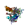
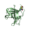
| Structure viewer | Molecule: MolmilJmol/JSmol |
|---|
- Downloads & links
-Download
| PDBx/mmCIF format | 5nyk.cif.gz | 69.6 KB | Display | PDBx/mmCIF format |
|---|---|---|---|---|
| PDB format | pdb5nyk.ent.gz | 49.9 KB | Display | PDB format |
| PDBx/mmJSON format | 5nyk.json.gz | Tree view | PDBx/mmJSON format | |
| Others |  Other downloads Other downloads |
-Validation report
| Arichive directory | https://data.pdbj.org/pub/pdb/validation_reports/ny/5nykftp://data.pdbj.org/pub/pdb/validation_reports/ny/5nyk | HTTPS FTP |
|---|
-Related structure data
| Related structure data |  5nylC  5nymC 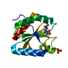 5nynC  5nyoC  1ep7S C: citing same article ( S: Starting model for refinement |
|---|---|
| Similar structure data |

-Links
 PDBj
PDBj

- Assembly
| Deposited unit | 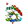
| ||||||||
|---|---|---|---|---|---|---|---|---|---|
| 1 |
| ||||||||
| Unit cell |
|
-Components
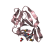
| #1: Protein | Mass: 14190.775 Da / Num. of mol.: 1 Source method: isolated from a genetically manipulated source Source: (gene. exp.) Populus tremula x Populus tremuloides (plant)Plasmid: pET3d / Details (production host): T7 expression strain / Production host:  Escherichia coli BL21(DE3) (bacteria) / References: UniProt: I0BZV0 Escherichia coli BL21(DE3) (bacteria) / References: UniProt: I0BZV0 | ||
|---|---|---|---|
| #2: Chemical | ChemComp-CL / Chloride  Mass: 35.453 Da / Num. of mol.: 1 / Source method: obtained synthetically / Formula: Cl Mass: 35.453 Da / Num. of mol.: 1 / Source method: obtained synthetically / Formula: Cl | ||
| #3: Chemical | ChemComp-K /   Mass: 39.098 Da / Num. of mol.: 1 / Source method: obtained synthetically / Formula: K Mass: 39.098 Da / Num. of mol.: 1 / Source method: obtained synthetically / Formula: K | ||
| #4: Chemical | Phosphate  Mass: 94.971 Da / Num. of mol.: 2 / Source method: obtained synthetically / Formula: PO4 Mass: 94.971 Da / Num. of mol.: 2 / Source method: obtained synthetically / Formula: PO4#5: Water | ChemComp-HOH / | Water Mass: 18.015 Da / Num. of mol.: 300 / Source method: isolated from a natural source / Formula: H2O Mass: 18.015 Da / Num. of mol.: 300 / Source method: isolated from a natural source / Formula: H2O |
-Experimental details
-Experiment
| Experiment | Method: X-RAY DIFFRACTION / Number of used crystals: 1 |
|---|
- Sample preparation
| Crystal | Density Matthews: 2.6 Å3/Da / Density % sol: 52.4 % |
|---|---|
| Crystal grow | Temperature: 291 K / Method: vapor diffusion, sitting drop / pH: 8 / Details: 0.4M NaH2PO4, 1.6M K2PO4, 0.2M NaCl, imidazole |
-Data collection
| Diffraction | Mean temperature: 110 K |
|---|---|
| Diffraction source | Source: SYNCHROTRON / Site: SOLEIL / Beamline: PROXIMA 1 / Wavelength: 0.82656 Å |
| Detector | Type: DECTRIS PILATUS 6M / Detector: PIXEL / Date: Aug 11, 2011 |
| Radiation | Monochromator: Si(111) / Protocol: SINGLE WAVELENGTH / Monochromatic (M) / Laue (L): M / Scattering type: x-ray |
| Radiation wavelength | Wavelength: 0.82656 Å / Relative weight: 1 |
| Reflection | Resolution: 1.05→39.4 Å / Num. obs: 66294 / % possible obs: 95.6 % / Redundancy: 13.5 % / Biso Wilson estimate: 8.16 Å2 / CC1/2: 0.999 / Rmerge(I) obs: 0.069 / Rpim(I) all: 0.28 / Net I/σ(I): 19.4 |
| Reflection shell | Resolution: 1.05→1.07 Å / Redundancy: 13.2 % / Rmerge(I) obs: 0.731 / Mean I/σ(I) obs: 3.4 / Num. unique obs: 3045 / CC1/2: 0.883 / Rpim(I) all: 0.295 / % possible all: 89.9 |
- Processing
| Software |
| ||||||||||||||||||||||||||||||||||||||||||||||||||||||||||||||||||||||||||||||||||||||||||||||||||||||||||||||||||
|---|---|---|---|---|---|---|---|---|---|---|---|---|---|---|---|---|---|---|---|---|---|---|---|---|---|---|---|---|---|---|---|---|---|---|---|---|---|---|---|---|---|---|---|---|---|---|---|---|---|---|---|---|---|---|---|---|---|---|---|---|---|---|---|---|---|---|---|---|---|---|---|---|---|---|---|---|---|---|---|---|---|---|---|---|---|---|---|---|---|---|---|---|---|---|---|---|---|---|---|---|---|---|---|---|---|---|---|---|---|---|---|---|---|---|---|
| Refinement | Method to determine structure: MOLECULAR REPLACEMENT Starting model: 1EP7 Resolution: 1.05→15.37 Å / Cor.coef. Fo:Fc: 0.967 / Cor.coef. Fo:Fc free: 0.9673 / SU R Cruickshank DPI: 0.025 / Cross valid method: THROUGHOUT / σ(F): 0 / SU R Blow DPI: 0.028 / SU Rfree Blow DPI: 0.028 / SU Rfree Cruickshank DPI: 0.024
| ||||||||||||||||||||||||||||||||||||||||||||||||||||||||||||||||||||||||||||||||||||||||||||||||||||||||||||||||||
| Displacement parameters | Biso mean: 13.4 Å2
| ||||||||||||||||||||||||||||||||||||||||||||||||||||||||||||||||||||||||||||||||||||||||||||||||||||||||||||||||||
| Refine analyze | Luzzati coordinate error obs: 0.107 Å | ||||||||||||||||||||||||||||||||||||||||||||||||||||||||||||||||||||||||||||||||||||||||||||||||||||||||||||||||||
| Refinement step | Cycle: LAST / Resolution: 1.05→15.37 Å
| ||||||||||||||||||||||||||||||||||||||||||||||||||||||||||||||||||||||||||||||||||||||||||||||||||||||||||||||||||
| Refine LS restraints |
| ||||||||||||||||||||||||||||||||||||||||||||||||||||||||||||||||||||||||||||||||||||||||||||||||||||||||||||||||||
| LS refinement shell | Resolution: 1.05→1.08 Å / Total num. of bins used: 20
|
