 Movie
Movie Controller


+ Open data

- Basic information
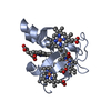
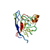
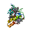
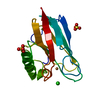
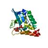
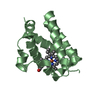
| Entry | Database: PDB / ID: 2cth | |||||||||
|---|---|---|---|---|---|---|---|---|---|---|
| Title | CYTOCHROME C3 FROM DESULFOVIBRIO VULGARIS HILDENBOROUGH | |||||||||
 Components Components | CYTOCHROME C3 | |||||||||
 Keywords Keywords |  ELECTRON TRANSPORT ELECTRON TRANSPORT | |||||||||
| Function / homology |  Function and homology informationanaerobic respiration / periplasmic space / electron transfer activity / heme binding / metal ion binding Function and homology informationanaerobic respiration / periplasmic space / electron transfer activity / heme binding / metal ion bindingSimilarity search - Function | |||||||||
| Biological species |  Desulfovibrio vulgaris subsp. vulgaris str. Hildenborough (bacteria) Desulfovibrio vulgaris subsp. vulgaris str. Hildenborough (bacteria) | |||||||||
| Method | X-RAY DIFFRACTION / SYNCHROTRON / MOLECULAR REPLACEMENT / Resolution: 1.67 Å | |||||||||
 Authors Authors | Simoes, P. / Matias, P.M. / Morais, J. / Wilson, K. / Dauter, Z. / Carrondo, M.A. | |||||||||
 Citation Citation | Journal: Inorg.Chim.Acta. / Year: 1998 Title: Refinement of the Three-Dimensional Structures of Cytochromes C3 from Desulfovibrio Vulgaris Hildenborough at 1.67 Angstrom Resolution and from Desulfovibrio Desulfuricans Atcc 27774 at 1.6 Angstrom Resolution Authors: Simoes, P. / Matias, P.M. / Morais, J. / Wilson, K. / Dauter, Z. / Carrondo, M.A. #1: Journal: J.Mol.Biol. / Year: 1993Title: Structure Analysis of Cytochrome C3 from Desulfovibrio Vulgaris Hildenborough at 1.9 A Resolution Authors: Matias, P.M. / Frazao, C. / Morais, J. / Coll, M. / Carrondo, M.A. | |||||||||
| History |
|
- Structure visualization
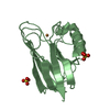
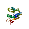
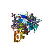
| Structure viewer | Molecule: MolmilJmol/JSmol |
|---|
- Downloads & links
-Download
| PDBx/mmCIF format | 2cth.cif.gz | 62.1 KB | Display | PDBx/mmCIF format |
|---|---|---|---|---|
| PDB format | pdb2cth.ent.gz | 50.5 KB | Display | PDB format |
| PDBx/mmJSON format | 2cth.json.gz | Tree view | PDBx/mmJSON format | |
| Others |  Other downloads Other downloads |
-Validation report
| Arichive directory | https://data.pdbj.org/pub/pdb/validation_reports/ct/2cthftp://data.pdbj.org/pub/pdb/validation_reports/ct/2cth | HTTPS FTP |
|---|
-Related structure data
| Related structure data | 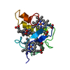 2cdvS S: Starting model for refinement |
|---|---|
| Similar structure data |

-Links
 PDBj
PDBj
- Assembly
| Deposited unit | 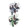
| ||||||||
|---|---|---|---|---|---|---|---|---|---|
| 1 | 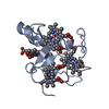
| ||||||||
| 2 | 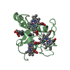
| ||||||||
| Unit cell |
|
-Components
| #1: Protein | Mass: 11687.463 Da / Num. of mol.: 2 / Source method: isolated from a natural source Source: (natural) Desulfovibrio vulgaris subsp. vulgaris str. Hildenborough (bacteria)Species: Desulfovibrio vulgaris / Strain: HILDENBOROUGH / References: UniProt: P00131#2: Chemical | ChemComp-HEM / Heme B  Mass: 616.487 Da / Num. of mol.: 8 / Source method: obtained synthetically / Formula: C34H32FeN4O4 Mass: 616.487 Da / Num. of mol.: 8 / Source method: obtained synthetically / Formula: C34H32FeN4O4#3: Water | ChemComp-HOH / | Water Mass: 18.015 Da / Num. of mol.: 124 / Source method: isolated from a natural source / Formula: H2O Mass: 18.015 Da / Num. of mol.: 124 / Source method: isolated from a natural source / Formula: H2O |
|---|
-Experimental details
-Experiment
| Experiment | Method: X-RAY DIFFRACTION / Number of used crystals: 1 |
|---|
- Sample preparation
| Crystal | Density Matthews: 2.84 Å3/Da / Density % sol: 56.75 % |
|---|---|
| Crystal grow | pH: 5.5 Details: PROTEIN WAS CRYSTALLIZED FROM 75% (V/V) ETHANOL AND 0.05 M SODIUM ACETATE (PH 5.5) |
-Data collection
| Diffraction | Mean temperature: 295 K |
|---|---|
| Diffraction source | Source: SYNCHROTRON / Site: EMBL/DESY, HAMBURG  / Beamline: X31 / Wavelength: 0.92 / Beamline: X31 / Wavelength: 0.92 |
| Detector | Type: MARRESEARCH / Detector: IMAGE PLATE / Date: Nov 1, 1993 / Details: MIRROR |
| Radiation | Monochromator: SI(111) / Monochromatic (M) / Laue (L): M / Scattering type: x-ray |
| Radiation wavelength | Wavelength: 0.92 Å / Relative weight: 1 |
| Reflection | Resolution: 1.67→25.3 Å / Num. obs: 28861 / % possible obs: 94.3 % / Observed criterion σ(I): 0 / Redundancy: 8.1 % / Rmerge(I) obs: 0.079 / Rsym value: 0.079 / Net I/σ(I): 8.1 |
| Reflection shell | Resolution: 1.67→1.71 Å / Redundancy: 4 % / Rmerge(I) obs: 0.265 / Mean I/σ(I) obs: 2.8 / Rsym value: 0.265 / % possible all: 89.6 |
- Processing
| Software |
| |||||||||||||||||||||||||||||||||
|---|---|---|---|---|---|---|---|---|---|---|---|---|---|---|---|---|---|---|---|---|---|---|---|---|---|---|---|---|---|---|---|---|---|---|
| Refinement | Method to determine structure: MOLECULAR REPLACEMENT Starting model: PDB ENTRY 2CDV Resolution: 1.67→8 Å / Num. parameters: 8266 / Num. restraintsaints: 30043 / Cross valid method: FREE R-VALUE / σ(F): 1
| |||||||||||||||||||||||||||||||||
| Refinement step | Cycle: LAST / Resolution: 1.67→8 Å
| |||||||||||||||||||||||||||||||||
| Refine LS restraints |
|
