 Movie
Movie Controller

[English] 日本語
 Yorodumi
- PDB-5ntt: Crystal structure of human Mps1 (TTK) C604Y mutant in complex wit... -
+ Open data

- Basic information
| Entry | Database: PDB / ID: 5ntt | ||||||||||||
|---|---|---|---|---|---|---|---|---|---|---|---|---|---|
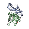
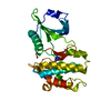
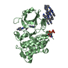
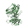
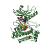
| Title | Crystal structure of human Mps1 (TTK) C604Y mutant in complex with NMS-P715 | ||||||||||||
 Components Components | Dual specificity protein kinase TTK | ||||||||||||
 Keywords Keywords |  TRANSFERASE / Mps1 / TTK / kinase / inhibitor / NMS-P715 / mutant / C604Y TRANSFERASE / Mps1 / TTK / kinase / inhibitor / NMS-P715 / mutant / C604Y | ||||||||||||
| Function / homology |  Function and homology information Function and homology informationprotein localization to meiotic spindle midzone / meiotic spindle assembly checkpoint signaling / kinetochore binding / female meiosis chromosome segregation / protein localization to kinetochore / dual-specificity kinase / spindle organization / mitotic spindle assembly checkpoint signaling / protein serine/threonine/tyrosine kinase activity / mitotic spindle organization ...protein localization to meiotic spindle midzone / meiotic spindle assembly checkpoint signaling / kinetochore binding / female meiosis chromosome segregation / protein localization to kinetochore / dual-specificity kinase / spindle organization / mitotic spindle assembly checkpoint signaling / protein serine/threonine/tyrosine kinase activity / mitotic spindle organization / chromosome segregation / kinetochore / spindle / protein tyrosine kinase activity / phosphorylation / protein serine kinase activity / protein serine/threonine kinase activity / positive regulation of cell population proliferation / ATP binding / membrane / identical protein binding / nucleus / cytoplasmSimilarity search - Function | ||||||||||||
| Biological species |  Homo sapiens (human) Homo sapiens (human) | ||||||||||||
| Method | X-RAY DIFFRACTION / SYNCHROTRON / MOLECULAR REPLACEMENT / molecular replacement / Resolution: 2.75 Å | ||||||||||||
 Authors Authors | Hiruma, Y. / Joosten, R.P. / Perrakis, A. | ||||||||||||
| Funding support |  Netherlands, 3items Netherlands, 3items
| ||||||||||||
 Citation Citation | Journal: J. Biol. Chem. / Year: 2017 Title: Understanding inhibitor resistance in Mps1 kinase through novel biophysical assays and structures. Authors: Hiruma, Y. / Koch, A. / Hazraty, N. / Tsakou, F. / Medema, R.H. / Joosten, R.P. / Perrakis, A. | ||||||||||||
| History |
|
- Structure visualization
| Structure viewer | Molecule: MolmilJmol/JSmol |
|---|
- Downloads & links
-Download
| PDBx/mmCIF format | 5ntt.cif.gz | 133.9 KB | Display | PDBx/mmCIF format |
|---|---|---|---|---|
| PDB format | pdb5ntt.ent.gz | 102.7 KB | Display | PDB format |
| PDBx/mmJSON format | 5ntt.json.gz | Tree view | PDBx/mmJSON format | |
| Others |  Other downloads Other downloads |
-Validation report
| Arichive directory | https://data.pdbj.org/pub/pdb/validation_reports/nt/5nttftp://data.pdbj.org/pub/pdb/validation_reports/nt/5ntt | HTTPS FTP |
|---|
-Related structure data
| Related structure data | 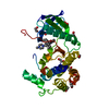 5mrbSC 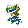 5o91C S: Starting model for refinement C: citing same article ( |
|---|---|
| Similar structure data |


-Links
 PDBj
PDBj- Assembly
| Deposited unit | 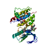
| ||||||||
|---|---|---|---|---|---|---|---|---|---|
| 1 |
| ||||||||
| Unit cell |
|
-Components
| #1: Protein | Mass: 33388.172 Da / Num. of mol.: 1 / Mutation: C604Y Source method: isolated from a genetically manipulated source Source: (gene. exp.) Homo sapiens (human) / Gene: TTK, MPS1, MPS1L1 / Production host:  Escherichia coli (E. coli) / References: UniProt: P33981, dual-specificity kinase Escherichia coli (E. coli) / References: UniProt: P33981, dual-specificity kinase | ||
|---|---|---|---|
| #2: Chemical | ChemComp-SVE /   Mass: 676.731 Da / Num. of mol.: 1 / Source method: obtained synthetically / Formula: C35H39F3N8O3 Mass: 676.731 Da / Num. of mol.: 1 / Source method: obtained synthetically / Formula: C35H39F3N8O3 | ||
| #3: Chemical | Ethylene glycol  Mass: 62.068 Da / Num. of mol.: 2 / Source method: obtained synthetically / Formula: C2H6O2 Mass: 62.068 Da / Num. of mol.: 2 / Source method: obtained synthetically / Formula: C2H6O2#4: Water | ChemComp-HOH / | Water Mass: 18.015 Da / Num. of mol.: 32 / Source method: isolated from a natural source / Formula: H2O Mass: 18.015 Da / Num. of mol.: 32 / Source method: isolated from a natural source / Formula: H2O |
-Experimental details
-Experiment
| Experiment | Method: X-RAY DIFFRACTION / Number of used crystals: 1 |
|---|
- Sample preparation
| Crystal | Density Matthews: 3.44 Å3/Da / Density % sol: 64.26 % |
|---|---|
| Crystal grow | Temperature: 291 K / Method: vapor diffusion, sitting drop / pH: 7.4 Details: 10.5% (w/v) PEG 350 MME, 10 mM MgCl2, and 100 mM Tris/HCl PH range: 7.5-9 |
-Data collection
| Diffraction | Mean temperature: 100 K | |||||||||||||||||||||
|---|---|---|---|---|---|---|---|---|---|---|---|---|---|---|---|---|---|---|---|---|---|---|
| Diffraction source | Source: SYNCHROTRON / Site: ESRF  / Beamline: ID29 / Wavelength: 1.07227 Å / Beamline: ID29 / Wavelength: 1.07227 Å | |||||||||||||||||||||
| Detector | Type: DECTRIS PILATUS 6M-F / Detector: PIXEL / Date: Mar 20, 2017 | |||||||||||||||||||||
| Radiation | Monochromator: Si (111) / Protocol: SINGLE WAVELENGTH / Monochromatic (M) / Laue (L): M / Scattering type: x-ray | |||||||||||||||||||||
| Radiation wavelength | Wavelength: 1.07227 Å / Relative weight: 1 | |||||||||||||||||||||
| Reflection | Resolution: 2.75→41.65 Å / Num. obs: 12326 / % possible obs: 100 % / Redundancy: 5.9 % / CC1/2: 0.996 / Rmerge(I) obs: 0.15 / Rpim(I) all: 0.067 / Rrim(I) all: 0.165 / Net I/σ(I): 9.6 / Num. measured all: 72378 / Scaling rejects: 0 | |||||||||||||||||||||
| Reflection shell | Diffraction-ID: 1
|
-Phasing
| Phasing | Method: molecular replacement | |||||||||
|---|---|---|---|---|---|---|---|---|---|---|
| Phasing MR | Model details: Phaser MODE: MR_AUTO
|
- Processing
| Software |
| ||||||||||||||||||||||||||||||||||||||||||||||||||||||||||||||||||||||||||||||||||||||||||||||||||||||||||||||||||||||||||||||||||||||||||||||||||||||||||||||||||||||||
|---|---|---|---|---|---|---|---|---|---|---|---|---|---|---|---|---|---|---|---|---|---|---|---|---|---|---|---|---|---|---|---|---|---|---|---|---|---|---|---|---|---|---|---|---|---|---|---|---|---|---|---|---|---|---|---|---|---|---|---|---|---|---|---|---|---|---|---|---|---|---|---|---|---|---|---|---|---|---|---|---|---|---|---|---|---|---|---|---|---|---|---|---|---|---|---|---|---|---|---|---|---|---|---|---|---|---|---|---|---|---|---|---|---|---|---|---|---|---|---|---|---|---|---|---|---|---|---|---|---|---|---|---|---|---|---|---|---|---|---|---|---|---|---|---|---|---|---|---|---|---|---|---|---|---|---|---|---|---|---|---|---|---|---|---|---|---|---|---|---|
| Refinement | Method to determine structure: MOLECULAR REPLACEMENT Starting model: 5MRB Resolution: 2.75→41.65 Å / Cor.coef. Fo:Fc: 0.951 / Cor.coef. Fo:Fc free: 0.923 / Matrix type: sparse / SU B: 29.224 / SU ML: 0.252 / Cross valid method: THROUGHOUT / σ(F): 0 / ESU R: 0.567 / ESU R Free: 0.288 Details: HYDROGENS HAVE BEEN ADDED IN THE RIDING POSITIONS U VALUES : WITH TLS ADDED
| ||||||||||||||||||||||||||||||||||||||||||||||||||||||||||||||||||||||||||||||||||||||||||||||||||||||||||||||||||||||||||||||||||||||||||||||||||||||||||||||||||||||||
| Solvent computation | Ion probe radii: 0.9 Å / Shrinkage radii: 0.9 Å / VDW probe radii: 1 Å | ||||||||||||||||||||||||||||||||||||||||||||||||||||||||||||||||||||||||||||||||||||||||||||||||||||||||||||||||||||||||||||||||||||||||||||||||||||||||||||||||||||||||
| Displacement parameters | Biso max: 179.74 Å2 / Biso mean: 73.976 Å2 / Biso min: 42.43 Å2
| ||||||||||||||||||||||||||||||||||||||||||||||||||||||||||||||||||||||||||||||||||||||||||||||||||||||||||||||||||||||||||||||||||||||||||||||||||||||||||||||||||||||||
| Refinement step | Cycle: final / Resolution: 2.75→41.65 Å
| ||||||||||||||||||||||||||||||||||||||||||||||||||||||||||||||||||||||||||||||||||||||||||||||||||||||||||||||||||||||||||||||||||||||||||||||||||||||||||||||||||||||||
| Refine LS restraints |
| ||||||||||||||||||||||||||||||||||||||||||||||||||||||||||||||||||||||||||||||||||||||||||||||||||||||||||||||||||||||||||||||||||||||||||||||||||||||||||||||||||||||||
| LS refinement shell | Refine-ID: X-RAY DIFFRACTION / Total num. of bins used: 20
| ||||||||||||||||||||||||||||||||||||||||||||||||||||||||||||||||||||||||||||||||||||||||||||||||||||||||||||||||||||||||||||||||||||||||||||||||||||||||||||||||||||||||
| Refinement TLS params. | Method: refined / Origin x: -4.3213 Å / Origin y: -21.4849 Å / Origin z: -17.1382 Å
|
