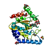
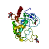
 Movie
Movie Controller


+ Open data

- Basic information
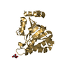
| Entry | Database: PDB / ID: 5nnh | ||||||
|---|---|---|---|---|---|---|---|
| Title | KSHV uracil-DNA glycosylase, apo form | ||||||
 Components Components | Uracil-DNA glycosylase | ||||||
 Keywords Keywords | HYDROLASE / Uracil-DNA glycosylase | ||||||
| Function / homology |  Function and homology informationbase-excision repair, AP site formation via deaminated base removal / uracil-DNA glycosylase / uracil DNA N-glycosylase activity / base-excision repair / host cell nucleus Function and homology informationbase-excision repair, AP site formation via deaminated base removal / uracil-DNA glycosylase / uracil DNA N-glycosylase activity / base-excision repair / host cell nucleusSimilarity search - Function | ||||||
| Biological species |   Human herpesvirus 8 Human herpesvirus 8 | ||||||
| Method | X-RAY DIFFRACTION / SYNCHROTRON / MOLECULAR REPLACEMENT / Resolution: 2.2 Å | ||||||
 Authors Authors | Earl, C. / Bagneris, C. / Cole, A.R. / Barrett, T. / Savva, R. | ||||||
| Funding support |  United Kingdom, 1items United Kingdom, 1items
| ||||||
 Citation Citation | Journal: Nucleic Acids Res. / Year: 2018 Title: A structurally conserved motif in gamma-herpesvirus uracil-DNA glycosylases elicits duplex nucleotide-flipping. Authors: Earl, C. / Bagneris, C. / Zeman, K. / Cole, A. / Barrett, T. / Savva, R. | ||||||
| History |
|
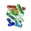
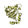
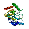
- Structure visualization
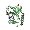
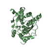
| Structure viewer | Molecule: MolmilJmol/JSmol |
|---|
- Downloads & links
-Download
| PDBx/mmCIF format | 5nnh.cif.gz | 105.3 KB | Display | PDBx/mmCIF format |
|---|---|---|---|---|
| PDB format | pdb5nnh.ent.gz | 79.4 KB | Display | PDB format |
| PDBx/mmJSON format | 5nnh.json.gz | Tree view | PDBx/mmJSON format | |
| Others |  Other downloads Other downloads |
-Validation report
| Arichive directory | https://data.pdbj.org/pub/pdb/validation_reports/nn/5nnhftp://data.pdbj.org/pub/pdb/validation_reports/nn/5nnh | HTTPS FTP |
|---|
-Related structure data
| Related structure data | 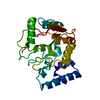 5nn7C  5nnuC 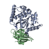 2j8xS C: citing same article ( S: Starting model for refinement |
|---|---|
| Similar structure data |

-Links
 PDBj
PDBj- Assembly
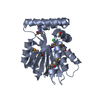
| Deposited unit | 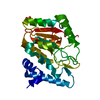
| ||||||||
|---|---|---|---|---|---|---|---|---|---|
| 1 |
| ||||||||
| Unit cell |
|
-Components
| #1: Protein | / UDG / UNG Mass: 27226.174 Da / Num. of mol.: 1 Source method: isolated from a genetically manipulated source Source: (gene. exp.) Human herpesvirus 8 / Gene: ORF46 / Plasmid: pRSET-C / Production host:  Escherichia coli (E. coli) / Strain (production host): T7 Express lysY/Iq Escherichia coli (E. coli) / Strain (production host): T7 Express lysY/IqReferences: UniProt: Q76RG8, UniProt: F5HFA1*PLUS, uracil-DNA glycosylase |
|---|---|
| #2: Chemical | ChemComp-SO4 / Sulfate  Mass: 96.063 Da / Num. of mol.: 1 / Source method: isolated from a natural source / Formula: SO4 Mass: 96.063 Da / Num. of mol.: 1 / Source method: isolated from a natural source / Formula: SO4 |
| #3: Water | ChemComp-HOH / Water Mass: 18.015 Da / Num. of mol.: 80 / Source method: isolated from a natural source / Formula: H2O Mass: 18.015 Da / Num. of mol.: 80 / Source method: isolated from a natural source / Formula: H2O |
-Experimental details
-Experiment
| Experiment | Method: X-RAY DIFFRACTION / Number of used crystals: 1 |
|---|
- Sample preparation
| Crystal | Density Matthews: 2.3 Å3/Da / Density % sol: 45.1 % |
|---|---|
| Crystal grow | Temperature: 289 K / Method: vapor diffusion, sitting drop / pH: 6.5 Details: 0.1 M bis-tris, 0.2 M lithium sulfate monohydrate, 25% PEG 3350 |
-Data collection
| Diffraction | Mean temperature: 100 K |
|---|---|
| Diffraction source | Source: SYNCHROTRON / Site: Diamond / Beamline: I24 / Wavelength: 0.96858 Å |
| Detector | Type: DECTRIS PILATUS3 S 6M / Detector: PIXEL / Date: Jan 14, 2017 |
| Radiation | Protocol: SINGLE WAVELENGTH / Monochromatic (M) / Laue (L): M / Scattering type: x-ray |
| Radiation wavelength | Wavelength: 0.96858 Å / Relative weight: 1 |
| Reflection | Resolution: 2.2→52.63 Å / Num. obs: 11700 / % possible obs: 100 % / Redundancy: 16.1 % / CC1/2: 0.996 / Rmerge(I) obs: 0.089 / Rpim(I) all: 0.204 / Net I/σ(I): 8.2 |
| Reflection shell | Resolution: 2.2→2.28 Å / Redundancy: 15 % / Rmerge(I) obs: 0.567 / Num. unique obs: 1027 / CC1/2: 0.858 / % possible all: 100 |
- Processing
| Software |
| |||||||||||||||||||||||||||||||||||||||||||||||||||||||||||||||||||||||||||
|---|---|---|---|---|---|---|---|---|---|---|---|---|---|---|---|---|---|---|---|---|---|---|---|---|---|---|---|---|---|---|---|---|---|---|---|---|---|---|---|---|---|---|---|---|---|---|---|---|---|---|---|---|---|---|---|---|---|---|---|---|---|---|---|---|---|---|---|---|---|---|---|---|---|---|---|---|
| Refinement | Method to determine structure: MOLECULAR REPLACEMENT Starting model: 2J8X Resolution: 2.2→52.51 Å / Cor.coef. Fo:Fc: 0.957 / Cor.coef. Fo:Fc free: 0.934 / Cross valid method: THROUGHOUT / σ(F): 0 / ESU R: 0.296 / ESU R Free: 0.22 Details: HYDROGENS HAVE BEEN ADDED IN THE RIDING POSITIONS U VALUES : WITH TLS ADDED
| |||||||||||||||||||||||||||||||||||||||||||||||||||||||||||||||||||||||||||
| Solvent computation | Ion probe radii: 0.8 Å / Shrinkage radii: 0.8 Å / VDW probe radii: 1.2 Å | |||||||||||||||||||||||||||||||||||||||||||||||||||||||||||||||||||||||||||
| Displacement parameters | Biso max: 94.91 Å2 / Biso mean: 50.2919 Å2 / Biso min: 28.75 Å2
| |||||||||||||||||||||||||||||||||||||||||||||||||||||||||||||||||||||||||||
| Refinement step | Cycle: LAST / Resolution: 2.2→52.51 Å
| |||||||||||||||||||||||||||||||||||||||||||||||||||||||||||||||||||||||||||
| Refine LS restraints |
| |||||||||||||||||||||||||||||||||||||||||||||||||||||||||||||||||||||||||||
| LS refinement shell | Resolution: 2.2→2.257 Å / Total num. of bins used: 20
| |||||||||||||||||||||||||||||||||||||||||||||||||||||||||||||||||||||||||||
| Refinement TLS params. | Method: refined / Origin x: -14.948 Å / Origin y: 10.559 Å / Origin z: 4.864 Å
|
