 Movie
Movie Controller

[English] 日本語
 Yorodumi
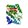
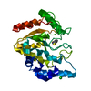
- PDB-1im8: Crystal structure of YecO from Haemophilus influenzae (HI0319), a... -
+ Open data

- Basic information
| Entry | Database: PDB / ID: 1im8 | ||||||
|---|---|---|---|---|---|---|---|
| Title | Crystal structure of YecO from Haemophilus influenzae (HI0319), a methyltransferase with a bound S-adenosylhomocysteine | ||||||
 Components Components | YecO | ||||||
 Keywords Keywords |  TRANSFERASE / methyltransferase / adenosylhomocysteine / structural genomics / hypothetical protein / Structure 2 Function Project / S2F TRANSFERASE / methyltransferase / adenosylhomocysteine / structural genomics / hypothetical protein / Structure 2 Function Project / S2F | ||||||
| Function / homology |  Function and homology informationTransferases; Transferring one-carbon groups; Carboxy- and carbamoyltransferases / carboxyl- or carbamoyltransferase activity / tRNA wobble uridine modification / S-adenosyl-L-methionine binding Function and homology informationTransferases; Transferring one-carbon groups; Carboxy- and carbamoyltransferases / carboxyl- or carbamoyltransferase activity / tRNA wobble uridine modification / S-adenosyl-L-methionine bindingSimilarity search - Function | ||||||
| Biological species |  Haemophilus influenzae Rd (bacteria) Haemophilus influenzae Rd (bacteria) | ||||||
| Method | X-RAY DIFFRACTION / SYNCHROTRON / MAD / Resolution: 2.2 Å | ||||||
 Authors Authors | Lim, K. / Zhang, H. / Tempczyk, A. / Bonander, N. / Toedt, J. / Howard, A. / Eisenstein, E. / Herzberg, O. / Structure 2 Function Project (S2F) | ||||||
 Citation Citation | Journal: Proteins / Year: 2001 Title: Crystal structure of YecO from Haemophilus influenzae (HI0319) reveals a methyltransferase fold and a bound S-adenosylhomocysteine. Authors: Lim, K. / Zhang, H. / Tempczyk, A. / Bonander, N. / Toedt, J. / Howard, A. / Eisenstein, E. / Herzberg, O. | ||||||
| History |
|
- Structure visualization
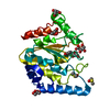
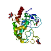
| Structure viewer | Molecule: MolmilJmol/JSmol |
|---|
- Downloads & links
-Download
| PDBx/mmCIF format | 1im8.cif.gz | 105.1 KB | Display | PDBx/mmCIF format |
|---|---|---|---|---|
| PDB format | pdb1im8.ent.gz | 85.6 KB | Display | PDB format |
| PDBx/mmJSON format | 1im8.json.gz | Tree view | PDBx/mmJSON format | |
| Others |  Other downloads Other downloads |
-Validation report
| Arichive directory | https://data.pdbj.org/pub/pdb/validation_reports/im/1im8ftp://data.pdbj.org/pub/pdb/validation_reports/im/1im8 | HTTPS FTP |
|---|
-Related structure data
| Similar structure data | |
|---|---|
| Other databases |
-Links
 PDBj
PDBj- Assembly
| Deposited unit | 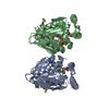
| ||||||||
|---|---|---|---|---|---|---|---|---|---|
| 1 | 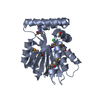
| ||||||||
| 2 | 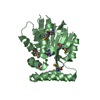
| ||||||||
| 3 | 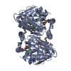
| ||||||||
| 4 | 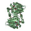
| ||||||||
| Unit cell |
|
-Components
| #1: Protein | Mass: 28015.070 Da / Num. of mol.: 2 Source method: isolated from a genetically manipulated source Source: (gene. exp.) Haemophilus influenzae Rd (bacteria) / Species: Haemophilus influenzae / Strain: KW20 / Gene: HI0319 / Production host: Escherichia coli (E. coli) / Strain (production host): B834(DE3)References: UniProt: P43985, Transferases; Transferring one-carbon groups; Methyltransferases#2: Chemical | Chloride  Mass: 35.453 Da / Num. of mol.: 2 / Source method: obtained synthetically / Formula: Cl Mass: 35.453 Da / Num. of mol.: 2 / Source method: obtained synthetically / Formula: Cl#3: Chemical | 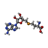  Mass: 431.306 Da / Num. of mol.: 2 / Source method: obtained synthetically / Formula: C14H20N6O5Se Mass: 431.306 Da / Num. of mol.: 2 / Source method: obtained synthetically / Formula: C14H20N6O5Se#4: Water | ChemComp-HOH / | Water Mass: 18.015 Da / Num. of mol.: 237 / Source method: isolated from a natural source / Formula: H2O Mass: 18.015 Da / Num. of mol.: 237 / Source method: isolated from a natural source / Formula: H2O |
|---|
-Experimental details
-Experiment
| Experiment | Method: X-RAY DIFFRACTION / Number of used crystals: 1 |
|---|
- Sample preparation
| Crystal | Density Matthews: 2.31 Å3/Da / Density % sol: 46.74 % | ||||||||||||||||||||||||||||||||||||||||||||||||
|---|---|---|---|---|---|---|---|---|---|---|---|---|---|---|---|---|---|---|---|---|---|---|---|---|---|---|---|---|---|---|---|---|---|---|---|---|---|---|---|---|---|---|---|---|---|---|---|---|---|
| Crystal grow | Temperature: 295 K / Method: vapor diffusion, hanging drop / pH: 7 Details: 20 % saturated ammonium sulfate, 100mM NaHepes with protein solution (10mg/ml in 150mM NaCl, 10mM NaHepes, pH 8.0, 1mM DTT and 0.5mM EDTA) , pH 7.0, VAPOR DIFFUSION, HANGING DROP, temperature 295K | ||||||||||||||||||||||||||||||||||||||||||||||||
| Crystal grow | *PLUS pH: 8 | ||||||||||||||||||||||||||||||||||||||||||||||||
| Components of the solutions | *PLUS
|
-Data collection
| Diffraction | Mean temperature: 100 K | ||||||||||||
|---|---|---|---|---|---|---|---|---|---|---|---|---|---|
| Diffraction source | Source: SYNCHROTRON / Site: APS  / Beamline: 17-ID / Wavelength: 0.9782, 0.9778, 0.9537 / Beamline: 17-ID / Wavelength: 0.9782, 0.9778, 0.9537 | ||||||||||||
| Detector | Type: BRUKER / Detector: CCD / Date: Jul 3, 1999 | ||||||||||||
| Radiation | Protocol: MAD / Monochromatic (M) / Laue (L): M / Scattering type: x-ray | ||||||||||||
| Radiation wavelength |
| ||||||||||||
| Reflection | Resolution: 2.2→20 Å / Num. all: 30031 / Num. obs: 30031 / % possible obs: 94 % / Observed criterion σ(F): 0 / Observed criterion σ(I): 0 / Redundancy: 3.2 % / Biso Wilson estimate: 28 Å2 / Rmerge(I) obs: 0.059 / Net I/σ(I): 13.2 | ||||||||||||
| Reflection shell | Resolution: 2.2→2.28 Å / Redundancy: 1.6 % / Rmerge(I) obs: 0.351 / % possible all: 74.6 | ||||||||||||
| Reflection | *PLUS Num. obs: 51819 / % possible obs: 93.7 % / Num. measured all: 174537 | ||||||||||||
| Reflection shell | *PLUS Highest resolution: 2.2 Å |
- Processing
| Software |
| ||||||||||||||||||||
|---|---|---|---|---|---|---|---|---|---|---|---|---|---|---|---|---|---|---|---|---|---|
| Refinement | Method to determine structure: MAD / Resolution: 2.2→20 Å / Cross valid method: THROUGHOUT / σ(F): 2 / σ(I): 2 / Stereochemistry target values: Engh & Huber
| ||||||||||||||||||||
| Refinement step | Cycle: LAST / Resolution: 2.2→20 Å
| ||||||||||||||||||||
| Refine LS restraints |
| ||||||||||||||||||||
| Software | *PLUS Name: CNS / Classification: refinement | ||||||||||||||||||||
| Refinement | *PLUS Highest resolution: 2.2 Å / Lowest resolution: 20 Å / σ(F): 2 / Rfactor obs: 0.192 / Rfactor Rfree: 0.257 | ||||||||||||||||||||
| Solvent computation | *PLUS | ||||||||||||||||||||
| Displacement parameters | *PLUS | ||||||||||||||||||||
| Refine LS restraints | *PLUS
| ||||||||||||||||||||
| LS refinement shell | *PLUS Rfactor Rfree: 0.315 / Rfactor obs: 0.253 |
