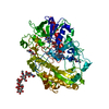
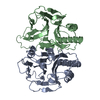
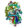
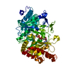
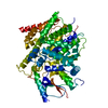
[English] 日本語
Yorodumi
- PDB-5nit: Glucose oxidase mutant A2 -

+
Open data


ID or keywords:

Loading...

-
Basic information

Entry
Database: PDB / ID: 5nit
TitleGlucose oxidase mutant A2
ComponentsGlucose oxidase
KeywordsOXIDOREDUCTASE / oxygen activation / His516 conformation
Function / homology
Function and homology information


glucose oxidase / glucose oxidase activity / flavin adenine dinucleotide binding / extracellular region
Similarity search - Function
Glucose Oxidase; domain 2 / Glucose Oxidase, domain 2 / Glucose Oxidase; domain 3 / Glucose Oxidase, domain 3 / Glucose Oxidase, domain 2 / GMC oxidoreductases signature 1. / GMC oxidoreductases signature 2. / Glucose-methanol-choline oxidoreductase / Glucose-methanol-choline oxidoreductase, N-terminal / GMC oxidoreductase ...Glucose Oxidase; domain 2 / Glucose Oxidase, domain 2 / Glucose Oxidase; domain 3 / Glucose Oxidase, domain 3 / Glucose Oxidase, domain 2 / GMC oxidoreductases signature 1. / GMC oxidoreductases signature 2. / Glucose-methanol-choline oxidoreductase / Glucose-methanol-choline oxidoreductase, N-terminal / GMC oxidoreductase / Glucose-methanol-choline oxidoreductase, C-terminal / GMC oxidoreductase / FAD/NAD(P)-binding domain / FAD/NAD(P)-binding domain / 3-Layer(bba) Sandwich / FAD/NAD(P)-binding domain superfamily / Few Secondary Structures / Irregular / 2-Layer Sandwich / Alpha Beta
Similarity search - Domain/homology
1,4-DIETHYLENE DIOXIDE / FLAVIN-ADENINE DINUCLEOTIDE / OXYGEN MOLECULE / Glucose oxidase
Similarity search - Component
Biological speciesAspergillus niger (mold)
MethodX-RAY DIFFRACTION / MOLECULAR REPLACEMENT / Resolution: 1.87 Å
AuthorsHoffmann, K.
Funding support Germany, 1items
OrganizationGrant numberCountry
Germany
CitationJournal: ACS Catal / Year: 2017
Title: Shuffling Active Site Substate Populations Affects Catalytic Activity: The Case of Glucose Oxidase.
Authors: Petrovic, D. / Frank, D. / Kamerlin, S.C.L. / Hoffmann, K. / Strodel, B.
History
DepositionMar 27, 2017Deposition site: PDBE / Processing site: PDBE
Revision 1.0Nov 15, 2017Provider: repository / Type: Initial release
Revision 1.1Jan 10, 2018Group: Database references / Category: citation
Item: _citation.journal_abbrev / _citation.journal_volume ..._citation.journal_abbrev / _citation.journal_volume / _citation.page_first / _citation.page_last / _citation.pdbx_database_id_PubMed / _citation.title
Revision 1.2Oct 16, 2019Group: Data collection / Category: reflns_shell
Revision 2.0Jul 29, 2020Group: Atomic model / Data collection ...Atomic model / Data collection / Derived calculations / Structure summary
Category: atom_site / atom_site_anisotrop ...atom_site / atom_site_anisotrop / chem_comp / diffrn_radiation_wavelength / entity / pdbx_branch_scheme / pdbx_chem_comp_identifier / pdbx_entity_branch / pdbx_entity_branch_descriptor / pdbx_entity_branch_link / pdbx_entity_branch_list / pdbx_entity_nonpoly / pdbx_nonpoly_scheme / pdbx_struct_assembly_gen / pdbx_struct_special_symmetry / struct_asym / struct_conn / struct_site / struct_site_gen
Item: _atom_site.B_iso_or_equiv / _atom_site.Cartn_x ..._atom_site.B_iso_or_equiv / _atom_site.Cartn_x / _atom_site.Cartn_y / _atom_site.Cartn_z / _atom_site.auth_asym_id / _atom_site.auth_atom_id / _atom_site.auth_comp_id / _atom_site.auth_seq_id / _atom_site.label_asym_id / _atom_site.label_atom_id / _atom_site.label_comp_id / _atom_site.label_entity_id / _atom_site.type_symbol / _atom_site_anisotrop.U[1][1] / _atom_site_anisotrop.U[1][2] / _atom_site_anisotrop.U[1][3] / _atom_site_anisotrop.U[2][2] / _atom_site_anisotrop.U[2][3] / _atom_site_anisotrop.U[3][3] / _atom_site_anisotrop.pdbx_auth_asym_id / _atom_site_anisotrop.pdbx_auth_atom_id / _atom_site_anisotrop.pdbx_auth_comp_id / _atom_site_anisotrop.pdbx_auth_seq_id / _atom_site_anisotrop.pdbx_label_asym_id / _atom_site_anisotrop.pdbx_label_atom_id / _atom_site_anisotrop.pdbx_label_comp_id / _atom_site_anisotrop.type_symbol / _chem_comp.name / _chem_comp.type / _pdbx_struct_assembly_gen.asym_id_list / _pdbx_struct_special_symmetry.label_asym_id / _struct_conn.pdbx_role / _struct_conn.ptnr1_auth_asym_id / _struct_conn.ptnr1_auth_seq_id / _struct_conn.ptnr1_label_asym_id / _struct_conn.ptnr2_auth_asym_id / _struct_conn.ptnr2_auth_seq_id / _struct_conn.ptnr2_label_asym_id
Description: Carbohydrate remediation / Provider: repository / Type: Remediation
Revision 2.1Jan 17, 2024Group: Data collection / Database references ...Data collection / Database references / Derived calculations / Refinement description / Structure summary
Category: chem_comp / chem_comp_atom ...chem_comp / chem_comp_atom / chem_comp_bond / database_2 / pdbx_initial_refinement_model / struct_conn
Item: _chem_comp.pdbx_synonyms / _database_2.pdbx_DOI ..._chem_comp.pdbx_synonyms / _database_2.pdbx_DOI / _database_2.pdbx_database_accession / _struct_conn.pdbx_leaving_atom_flag

-
Structure visualization

Structure viewerMolecule:
MolmilJmol/JSmol

Downloads & links

-
Assembly

Deposited unit
A: Glucose oxidase
hetero molecules


Theoretical massNumber of molelcules
Total (without water)67,13626
Polymers63,0821
Non-polymers4,05425
Water7,692427
1


  • Idetical with deposited unit
  • defined by author&software
  • Evidence: gel filtration
TypeNameSymmetry operationNumber
identity operation1_555x,y,z1
Buried area6930 Å2
ΔGint81 kcal/mol
Surface area21100 Å2
MethodPISA
Unit cell
Length a, b, c (Å)128.657, 128.657, 77.693
Angle α, β, γ (deg.)90.00, 90.00, 120.00
Int Tables number154
Space group name H-MP3221
Components on special symmetry positions
IDModelComponents
11A-1004-

HOH

-
Components

-
Protein , 1 types, 1 molecules A

#1: Protein Glucose oxidase / / Beta-D-glucose:oxygen 1-oxido-reductase / Glucose oxyhydrase / GOD


Mass: 63081.777 Da / Num. of mol.: 1 / Mutation: T30V I94V A162T R537K M556V
Source method: isolated from a genetically manipulated source
Details: mutant A2 / Source: (gene. exp.) Aspergillus niger (mold) / Gene: gox / Production host: Komagataella pastoris (fungus) / References: UniProt: P13006, glucose oxidase

-
Sugars , 2 types, 4 molecules

#2: Polysaccharide alpha-D-mannopyranose-(1-6)-beta-D-mannopyranose-(1-4)-2-acetamido-2-deoxy-beta-D-glucopyranose-(1- ...alpha-D-mannopyranose-(1-6)-beta-D-mannopyranose-(1-4)-2-acetamido-2-deoxy-beta-D-glucopyranose-(1-4)-2-acetamido-2-deoxy-beta-D-glucopyranose


Type: oligosaccharide / Mass: 748.682 Da / Num. of mol.: 1
Source method: isolated from a genetically manipulated source
DescriptorTypeProgram
DManpa1-6DManpb1-4DGlcpNAcb1-4DGlcpNAcb1-Glycam Condensed SequenceGMML 1.0
WURCS=2.0/3,4,3/[a2122h-1b_1-5_2*NCC/3=O][a1122h-1b_1-5][a1122h-1a_1-5]/1-1-2-3/a4-b1_b4-c1_c6-d1WURCSPDB2Glycan 1.1.0
[]{[(4+1)][b-D-GlcpNAc]{[(4+1)][b-D-GlcpNAc]{[(4+1)][b-D-Manp]{[(6+1)][a-D-Manp]{}}}}}LINUCSPDB-CARE
#5: Sugar ChemComp-NAG / 2-acetamido-2-deoxy-beta-D-glucopyranose / N-acetyl-beta-D-glucosamine / 2-acetamido-2-deoxy-beta-D-glucose / 2-acetamido-2-deoxy-D-glucose / 2-acetamido-2-deoxy-glucose / N-ACETYL-D-GLUCOSAMINE / N-Acetylglucosamine


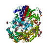
Type: D-saccharide, beta linking / Mass: 221.208 Da / Num. of mol.: 3
Source method: isolated from a genetically manipulated source
Formula: C8H15NO6
IdentifierTypeProgram
DGlcpNAcbCONDENSED IUPAC CARBOHYDRATE SYMBOLGMML 1.0
N-acetyl-b-D-glucopyranosamineCOMMON NAMEGMML 1.0
b-D-GlcpNAcIUPAC CARBOHYDRATE SYMBOLPDB-CARE 1.0
GlcNAcSNFG CARBOHYDRATE SYMBOLGMML 1.0

-
Non-polymers , 5 types, 448 molecules

#3: Chemical ChemComp-FAD / FLAVIN-ADENINE DINUCLEOTIDE / Flavin adenine dinucleotide


Mass: 785.550 Da / Num. of mol.: 1 / Source method: obtained synthetically / Formula: C27H33N9O15P2 / Comment: FAD*YM
#4: Chemical ChemComp-OXY / OXYGEN MOLECULE / Oxygen


Mass: 31.999 Da / Num. of mol.: 1 / Source method: obtained synthetically / Formula: O2
#6: Chemical
ChemComp-DIO / 1,4-DIETHYLENE DIOXIDE / 1,4-Dioxane


Mass: 88.105 Da / Num. of mol.: 18
Source method: isolated from a genetically manipulated source
Formula: C4H8O2
#7: Chemical ChemComp-EPE / 4-(2-HYDROXYETHYL)-1-PIPERAZINE ETHANESULFONIC ACID / HEPES / HEPES


Mass: 238.305 Da / Num. of mol.: 1 / Source method: obtained synthetically / Formula: C8H18N2O4S / Comment: pH buffer*YM
#8: Water ChemComp-HOH / water / Water


Mass: 18.015 Da / Num. of mol.: 427 / Source method: isolated from a natural source / Formula: H2O

-
Experimental details

-
Experiment

ExperimentMethod: X-RAY DIFFRACTION / Number of used crystals: 1

-
Sample preparation

CrystalDensity Matthews: 2.94 Å3/Da / Density % sol: 58.2 %
Crystal growTemperature: 280 K / Method: vapor diffusion, sitting drop / pH: 7 / Details: 100 mM HEPES 40% v/v dioxane

-
Data collection

DiffractionMean temperature: 100 K
Diffraction sourceSource: ROTATING ANODE / Type: ENRAF-NONIUS FR591 / Wavelength: 1.54 Å
DetectorType: MAR scanner 345 mm plate / Detector: IMAGE PLATE / Date: May 23, 2013
RadiationProtocol: SINGLE WAVELENGTH / Monochromatic (M) / Laue (L): M / Scattering type: x-ray
Radiation wavelengthWavelength: 1.54 Å / Relative weight: 1
ReflectionResolution: 1.86→45.27 Å / Num. obs: 61296 / % possible obs: 98.1 % / Redundancy: 8.2 % / Net I/σ(I): 12.6

-
Processing

Software
NameVersionClassification
REFMAC5.8.0158refinement
iMOSFLM7.2.1data reduction
SCALA6.5.019data scaling
PHASER7.0.20phasing
RefinementMethod to determine structure: MOLECULAR REPLACEMENT
Starting model: 1CF3
Resolution: 1.87→111.42 Å / Cor.coef. Fo:Fc: 0.944 / Cor.coef. Fo:Fc free: 0.919 / SU B: 5.997 / SU ML: 0.078 / Cross valid method: THROUGHOUT / ESU R: 0.117 / ESU R Free: 0.116 / Stereochemistry target values: MAXIMUM LIKELIHOOD / Details: HYDROGENS HAVE BEEN ADDED IN THE RIDING POSITIONS
RfactorNum. reflection% reflectionSelection details
Rfree0.20393 3243 5.3 %RANDOM
Rwork0.16472 ---
obs0.16678 58032 99.45 %-
Solvent computationIon probe radii: 0.8 Å / Shrinkage radii: 0.8 Å / VDW probe radii: 0.5 Å / Solvent model: BABINET MODEL WITH MASK
Displacement parametersBiso mean: 20.215 Å2
Baniso -1Baniso -2Baniso -3
1--0.04 Å2-0.02 Å2-0 Å2
2---0.04 Å20 Å2
3---0.13 Å2
Refinement stepCycle: 1 / Resolution: 1.87→111.42 Å
ProteinNucleic acidLigandSolventTotal
Num. atoms4430 0 270 427 5127
Refine LS restraints
Refine-IDTypeDev idealDev ideal targetNumber
X-RAY DIFFRACTIONr_bond_refined_d0.0190.0194873
X-RAY DIFFRACTIONr_bond_other_d0.0020.024304
X-RAY DIFFRACTIONr_angle_refined_deg1.9521.9936645
X-RAY DIFFRACTIONr_angle_other_deg1.115310037
X-RAY DIFFRACTIONr_dihedral_angle_1_deg6.7425599
X-RAY DIFFRACTIONr_dihedral_angle_2_deg35.69824.554213
X-RAY DIFFRACTIONr_dihedral_angle_3_deg11.9115685
X-RAY DIFFRACTIONr_dihedral_angle_4_deg14.8591521
X-RAY DIFFRACTIONr_chiral_restr0.1170.2738
X-RAY DIFFRACTIONr_gen_planes_refined0.0110.0215361
X-RAY DIFFRACTIONr_gen_planes_other0.0020.02959
X-RAY DIFFRACTIONr_nbd_refined
X-RAY DIFFRACTIONr_nbd_other
X-RAY DIFFRACTIONr_nbtor_refined
X-RAY DIFFRACTIONr_nbtor_other
X-RAY DIFFRACTIONr_xyhbond_nbd_refined
X-RAY DIFFRACTIONr_xyhbond_nbd_other
X-RAY DIFFRACTIONr_metal_ion_refined
X-RAY DIFFRACTIONr_metal_ion_other
X-RAY DIFFRACTIONr_symmetry_vdw_refined
X-RAY DIFFRACTIONr_symmetry_vdw_other
X-RAY DIFFRACTIONr_symmetry_hbond_refined
X-RAY DIFFRACTIONr_symmetry_hbond_other
X-RAY DIFFRACTIONr_symmetry_metal_ion_refined
X-RAY DIFFRACTIONr_symmetry_metal_ion_other
X-RAY DIFFRACTIONr_mcbond_it1.0481.5582343
X-RAY DIFFRACTIONr_mcbond_other1.0411.5562342
X-RAY DIFFRACTIONr_mcangle_it1.6642.3312932
X-RAY DIFFRACTIONr_mcangle_other1.6642.3342933
X-RAY DIFFRACTIONr_scbond_it1.6471.8452528
X-RAY DIFFRACTIONr_scbond_other1.6451.8442528
X-RAY DIFFRACTIONr_scangle_it
X-RAY DIFFRACTIONr_scangle_other2.5622.6693702
X-RAY DIFFRACTIONr_long_range_B_refined4.25919.8035755
X-RAY DIFFRACTIONr_long_range_B_other4.25919.8085756
X-RAY DIFFRACTIONr_rigid_bond_restr
X-RAY DIFFRACTIONr_sphericity_free
X-RAY DIFFRACTIONr_sphericity_bonded
LS refinement shellResolution: 1.868→1.916 Å / Total num. of bins used: 20
RfactorNum. reflection% reflection
Rfree0.244 191 -
Rwork0.254 4124 -
obs--95.85 %
Refinement TLS params.Method: refined / Origin x: 87.7078 Å / Origin y: 48.8579 Å / Origin z: 9.6658 Å
111213212223313233
T0.0101 Å2-0.0167 Å2-0.0008 Å2-0.0364 Å20.0013 Å2--0.0031 Å2
L0.3679 °20.0542 °20.044 °2-0.1125 °20.0185 °2--0.1685 °2
S-0.0057 Å °0.0561 Å °-0.014 Å °-0.005 Å °0.0164 Å °0.0108 Å °-0.0181 Å °0.0154 Å °-0.0107 Å °

+
About Yorodumi

-
News

-
Feb 9, 2022. New format data for meta-information of EMDB entries

New format data for meta-information of EMDB entries

  • Version 3 of the EMDB header file is now the official format.
  • The previous official version 1.9 will be removed from the archive.

Related info.:EMDB header

External links:wwPDB to switch to version 3 of the EMDB data model

-
Aug 12, 2020. Covid-19 info

Covid-19 info

URL: https://pdbj.org/emnavi/covid19.php

New page: Covid-19 featured information page in EM Navigator.

Related info.:Covid-19 info / Mar 5, 2020. Novel coronavirus structure data

+
Mar 5, 2020. Novel coronavirus structure data

Novel coronavirus structure data

Related info.:Yorodumi Speices / Aug 12, 2020. Covid-19 info

External links:COVID-19 featured content - PDBj / Molecule of the Month (242):Coronavirus Proteases

+
Jan 31, 2019. EMDB accession codes are about to change! (news from PDBe EMDB page)

EMDB accession codes are about to change! (news from PDBe EMDB page)

  • The allocation of 4 digits for EMDB accession codes will soon come to an end. Whilst these codes will remain in use, new EMDB accession codes will include an additional digit and will expand incrementally as the available range of codes is exhausted. The current 4-digit format prefixed with “EMD-” (i.e. EMD-XXXX) will advance to a 5-digit format (i.e. EMD-XXXXX), and so on. It is currently estimated that the 4-digit codes will be depleted around Spring 2019, at which point the 5-digit format will come into force.
  • The EM Navigator/Yorodumi systems omit the EMD- prefix.

Related info.:Q: What is EMD? / ID/Accession-code notation in Yorodumi/EM Navigator

External links:EMDB Accession Codes are Changing Soon! / Contact to PDBj

+
Jul 12, 2017. Major update of PDB

Major update of PDB

  • wwPDB released updated PDB data conforming to the new PDBx/mmCIF dictionary.
  • This is a major update changing the version number from 4 to 5, and with Remediation, in which all the entries are updated.
  • In this update, many items about electron microscopy experimental information are reorganized (e.g. em_software).
  • Now, EM Navigator and Yorodumi are based on the updated data.

External links:wwPDB Remediation / Enriched Model Files Conforming to OneDep Data Standards Now Available in the PDB FTP Archive

-
Yorodumi

Thousand views of thousand structures

  • Yorodumi is a browser for structure data from EMDB, PDB, SASBDB, etc.
  • This page is also the successor to EM Navigator detail page, and also detail information page/front-end page for Omokage search.
  • The word "yorodu" (or yorozu) is an old Japanese word meaning "ten thousand". "mi" (miru) is to see.

Related info.:EMDB / PDB / SASBDB / Comparison of 3 databanks / Yorodumi Search / Aug 31, 2016. New EM Navigator & Yorodumi / Yorodumi Papers / Jmol/JSmol / Function and homology information / Changes in new EM Navigator and Yorodumi

Read more