 Movie
Movie Controller

[English] 日本語
 Yorodumi
- PDB-5nig: Crystal structure of HLA-DRB1*04:01 with modified alpha-enolase p... -
+ Open data

- Basic information
| Entry | Database: PDB / ID: 5nig | |||||||||
|---|---|---|---|---|---|---|---|---|---|---|
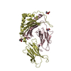
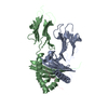
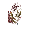
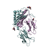
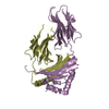
| Title | Crystal structure of HLA-DRB1*04:01 with modified alpha-enolase peptide 326-340 (arginine 327 to citrulline) | |||||||||
 Components Components |
| |||||||||
 Keywords Keywords |  IMMUNE SYSTEM / HLA-DR / enolase / arthritis / citrulline IMMUNE SYSTEM / HLA-DR / enolase / arthritis / citrulline | |||||||||
| Function / homology |  Function and homology information Function and homology informationnegative regulation of hypoxia-induced intrinsic apoptotic signaling pathway / Manipulation of host energy metabolism / positive regulation of muscle contraction / positive regulation of plasminogen activation / regulation of interleukin-4 production / regulation of interleukin-10 production / phosphopyruvate hydratase / phosphopyruvate hydratase complex / phosphopyruvate hydratase activity / positive regulation of T cell mediated immune response to tumor cell ...negative regulation of hypoxia-induced intrinsic apoptotic signaling pathway / Manipulation of host energy metabolism / positive regulation of muscle contraction / positive regulation of plasminogen activation / regulation of interleukin-4 production / regulation of interleukin-10 production / phosphopyruvate hydratase / phosphopyruvate hydratase complex / phosphopyruvate hydratase activity / positive regulation of T cell mediated immune response to tumor cell / myeloid dendritic cell antigen processing and presentation / antigen processing and presentation of endogenous peptide antigen via MHC class II / autolysosome membrane / regulation of T-helper cell differentiation / positive regulation of CD4-positive, CD25-positive, alpha-beta regulatory T cell differentiation / MHC class II receptor activity / positive regulation of CD4-positive, alpha-beta T cell activation / antigen processing and presentation of peptide or polysaccharide antigen via MHC class II / positive regulation of memory T cell differentiation / positive regulation of monocyte differentiation / CD4 receptor binding / Gluconeogenesis / nuclear outer membrane / positive regulation of kinase activity / inflammatory response to antigenic stimulus / M band / canonical glycolysis / Glycolysis / transport vesicle membrane / intermediate filament / polysaccharide binding / T-helper 1 type immune response / positive regulation of ATP biosynthetic process / Translocation of ZAP-70 to Immunological synapse / Phosphorylation of CD3 and TCR zeta chains / positive regulation of insulin secretion involved in cellular response to glucose stimulus / humoral immune response / macrophage differentiation / negative regulation of type II interferon production / Generation of second messenger molecules / immunological synapse / PD-1 signaling / epidermis development / detection of bacterium / T cell receptor binding / negative regulation of T cell proliferation / negative regulation of inflammatory response to antigenic stimulus / MHC class II antigen presentation / transcription corepressor binding / trans-Golgi network membrane / gluconeogenesis / lumenal side of endoplasmic reticulum membrane / glycolytic process / protein tetramerization / RNA polymerase II transcription regulatory region sequence-specific DNA binding / clathrin-coated endocytic vesicle membrane / response to virus / ER to Golgi transport vesicle membrane / negative regulation of cell growth / structural constituent of cytoskeleton / cognition / DNA-binding transcription repressor activity, RNA polymerase II-specific / peptide antigen assembly with MHC class II protein complex / MHC class II protein complex / positive regulation of T cell mediated cytotoxicity / peptide antigen binding / transcription corepressor activity / endocytic vesicle membrane / antigen processing and presentation of exogenous peptide antigen via MHC class II / Interferon gamma signaling / positive regulation of immune response / positive regulation of T cell activation / Downstream TCR signaling / GTPase binding / MHC class II protein complex binding / late endosome membrane / T cell receptor signaling pathway / cell cortex / early endosome membrane / positive regulation of canonical NF-kappaB signal transduction / adaptive immune response / positive regulation of MAPK cascade / positive regulation of viral entry into host cell / lysosome / positive regulation of ERK1 and ERK2 cascade / cadherin binding / immune response / positive regulation of protein phosphorylation / lysosomal membrane / external side of plasma membrane / Golgi membrane / negative regulation of DNA-templated transcription / positive regulation of DNA-templated transcription / negative regulation of transcription by RNA polymerase II / magnesium ion binding / cell surface / signal transduction / protein homodimerization activity / extracellular space / RNA bindingSimilarity search - Function | |||||||||
| Biological species |  Homo sapiens (human) Homo sapiens (human) | |||||||||
| Method | X-RAY DIFFRACTION / SYNCHROTRON / MOLECULAR REPLACEMENT / Resolution: 1.35 Å | |||||||||
 Authors Authors | Gerstner, C. / Dubnovitsky, A. | |||||||||
 Citation Citation | Journal: J. Autoimmun. / Year: 2018 Title: Memory T cells specific to citrullinated alpha-enolase are enriched in the rheumatic joint. Authors: Pieper, J. / Dubnovitsky, A. / Gerstner, C. / James, E.A. / Rieck, M. / Kozhukh, G. / Tandre, K. / Pellegrino, S. / Gebe, J.A. / Ronnblom, L. / Sandalova, T. / Kwok, W.W. / Klareskog, L. / ...Authors: Pieper, J. / Dubnovitsky, A. / Gerstner, C. / James, E.A. / Rieck, M. / Kozhukh, G. / Tandre, K. / Pellegrino, S. / Gebe, J.A. / Ronnblom, L. / Sandalova, T. / Kwok, W.W. / Klareskog, L. / Buckner, J.H. / Achour, A. / Malmstrom, V. | |||||||||
| History |
|
- Structure visualization
| Structure viewer | Molecule: MolmilJmol/JSmol |
|---|
- Downloads & links
-Download
| PDBx/mmCIF format | 5nig.cif.gz | 197.9 KB | Display | PDBx/mmCIF format |
|---|---|---|---|---|
| PDB format | pdb5nig.ent.gz | 157.2 KB | Display | PDB format |
| PDBx/mmJSON format | 5nig.json.gz | Tree view | PDBx/mmJSON format | |
| Others |  Other downloads Other downloads |
-Validation report
| Arichive directory | https://data.pdbj.org/pub/pdb/validation_reports/ni/5nigftp://data.pdbj.org/pub/pdb/validation_reports/ni/5nig | HTTPS FTP |
|---|
-Related structure data
| Related structure data |  5ni9SC S: Starting model for refinement C: citing same article ( |
|---|---|
| Similar structure data |


-Links
 PDBj
PDBj


- Assembly
| Deposited unit | 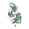
| ||||||||
|---|---|---|---|---|---|---|---|---|---|
| 1 |
| ||||||||
| Unit cell |
|
-Components
-HLA class II histocompatibility antigen, ... , 2 types, 2 molecules AB
| #1: Protein | Mass: 21911.750 Da / Num. of mol.: 1 Source method: isolated from a genetically manipulated source Source: (gene. exp.) Homo sapiens (human) / Gene: HLA-DRA, HLA-DRA1 / Production host:  Escherichia coli (E. coli) / References: UniProt: P01903 Escherichia coli (E. coli) / References: UniProt: P01903 |
|---|---|
| #2: Protein | Mass: 23102.672 Da / Num. of mol.: 1 Source method: isolated from a genetically manipulated source Source: (gene. exp.) Homo sapiens (human) / Gene: HLA-DRB1 / Production host: Escherichia coli (E. coli) / References: UniProt: P13760, UniProt: P01911*PLUS |
-Protein/peptide , 1 types, 1 molecules C
| #3: Protein/peptide | / 2-phospho-D-glycerate hydro-lyase / C-myc promoter-binding protein / Enolase 1 / MBP-1 / MPB-1 / ...2-phospho-D-glycerate hydro-lyase / C-myc promoter-binding protein / Enolase 1 / MBP-1 / MPB-1 / Non-neural enolase / NNE / Phosphopyruvate hydratase / Plasminogen-binding protein Mass: 1681.010 Da / Num. of mol.: 1 / Fragment: UNP Residues 326-340 / Source method: obtained synthetically / Source: (synth.) Homo sapiens (human) / References: UniProt: P06733, phosphopyruvate hydratase |
|---|
-Non-polymers , 4 types, 419 molecules 


| #4: Chemical | 2-Methyl-2,4-pentanediol Mass: 118.174 Da / Num. of mol.: 3 / Source method: obtained synthetically / Formula: C6H14O2 / Comment: precipitant*YM Mass: 118.174 Da / Num. of mol.: 3 / Source method: obtained synthetically / Formula: C6H14O2 / Comment: precipitant*YM#5: Chemical | ChemComp-URE / | Urea Mass: 60.055 Da / Num. of mol.: 1 / Source method: obtained synthetically / Formula: CH4N2O Mass: 60.055 Da / Num. of mol.: 1 / Source method: obtained synthetically / Formula: CH4N2O#6: Chemical | ChemComp-PGE / | Polyethylene glycol Mass: 150.173 Da / Num. of mol.: 1 / Source method: obtained synthetically / Formula: C6H14O4 Mass: 150.173 Da / Num. of mol.: 1 / Source method: obtained synthetically / Formula: C6H14O4#7: Water | ChemComp-HOH / | WaterMass: 18.015 Da / Num. of mol.: 414 / Source method: isolated from a natural source / Formula: H2O |
|---|
-Experimental details
-Experiment
| Experiment | Method: X-RAY DIFFRACTION / Number of used crystals: 1 |
|---|
- Sample preparation
| Crystal | Density Matthews: 2.52 Å3/Da / Density % sol: 51.24 % / Description: rod-like big crystals |
|---|---|
| Crystal grow | Temperature: 293 K / Method: vapor diffusion, hanging drop / pH: 6.5 Details: 0.1 M MES pH 6.5 10% (vol/vol) MPD 15% (vol/vol) PEG 3350 |
-Data collection
| Diffraction | Mean temperature: 100 K |
|---|---|
| Diffraction source | Source: SYNCHROTRON / Site: ESRF  / Beamline: MASSIF-1 / Wavelength: 0.966 Å / Beamline: MASSIF-1 / Wavelength: 0.966 Å |
| Detector | Type: DECTRIS PILATUS3 2M / Detector: PIXEL / Date: May 13, 2016 |
| Radiation | Protocol: SINGLE WAVELENGTH / Monochromatic (M) / Laue (L): M / Scattering type: x-ray |
| Radiation wavelength | Wavelength: 0.966 Å / Relative weight: 1 |
| Reflection | Resolution: 1.35→46.86 Å / Num. obs: 103630 / % possible obs: 98.9 % / Redundancy: 4.4 % / Biso Wilson estimate: 23.5 Å2 / CC1/2: 0.996 / Net I/σ(I): 9.9 |
- Processing
| Software |
| ||||||||||||||||||||||||||||||||||||||||||||||||||||||||||||||||||||||||||||||||||||||||||||||||||||||||||||||||||||||||||||||||||||||||||||||||||||||||||||||||||||||||||||||||||||||
|---|---|---|---|---|---|---|---|---|---|---|---|---|---|---|---|---|---|---|---|---|---|---|---|---|---|---|---|---|---|---|---|---|---|---|---|---|---|---|---|---|---|---|---|---|---|---|---|---|---|---|---|---|---|---|---|---|---|---|---|---|---|---|---|---|---|---|---|---|---|---|---|---|---|---|---|---|---|---|---|---|---|---|---|---|---|---|---|---|---|---|---|---|---|---|---|---|---|---|---|---|---|---|---|---|---|---|---|---|---|---|---|---|---|---|---|---|---|---|---|---|---|---|---|---|---|---|---|---|---|---|---|---|---|---|---|---|---|---|---|---|---|---|---|---|---|---|---|---|---|---|---|---|---|---|---|---|---|---|---|---|---|---|---|---|---|---|---|---|---|---|---|---|---|---|---|---|---|---|---|---|---|---|---|
| Refinement | Method to determine structure: MOLECULAR REPLACEMENT Starting model: 5NI9 Resolution: 1.35→46.86 Å / Cor.coef. Fo:Fc: 0.978 / Cor.coef. Fo:Fc free: 0.959 / SU B: 2.062 / SU ML: 0.036 / Cross valid method: THROUGHOUT / ESU R: 0.046 / ESU R Free: 0.047 / Details: HYDROGENS HAVE BEEN ADDED IN THE RIDING POSITIONS
| ||||||||||||||||||||||||||||||||||||||||||||||||||||||||||||||||||||||||||||||||||||||||||||||||||||||||||||||||||||||||||||||||||||||||||||||||||||||||||||||||||||||||||||||||||||||
| Solvent computation | Ion probe radii: 0.8 Å / Shrinkage radii: 0.8 Å / VDW probe radii: 1.2 Å | ||||||||||||||||||||||||||||||||||||||||||||||||||||||||||||||||||||||||||||||||||||||||||||||||||||||||||||||||||||||||||||||||||||||||||||||||||||||||||||||||||||||||||||||||||||||
| Displacement parameters | Biso mean: 22.68 Å2
| ||||||||||||||||||||||||||||||||||||||||||||||||||||||||||||||||||||||||||||||||||||||||||||||||||||||||||||||||||||||||||||||||||||||||||||||||||||||||||||||||||||||||||||||||||||||
| Refinement step | Cycle: 1 / Resolution: 1.35→46.86 Å
| ||||||||||||||||||||||||||||||||||||||||||||||||||||||||||||||||||||||||||||||||||||||||||||||||||||||||||||||||||||||||||||||||||||||||||||||||||||||||||||||||||||||||||||||||||||||
| Refine LS restraints |
|