 Movie
Movie Controller

[English] 日本語
 Yorodumi
- PDB-5nfj: Crystal structure of the methyltransferase subunit of human mitoc... -
+ Open data

- Basic information
| Entry | Database: PDB / ID: 5nfj | ||||||
|---|---|---|---|---|---|---|---|
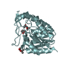
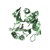
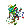
| Title | Crystal structure of the methyltransferase subunit of human mitochondrial Ribonuclease P (MRPP1) bound to S-adenosyl-methionine (SAM) | ||||||
 Components Components | Mitochondrial ribonuclease P protein 1 | ||||||
 Keywords Keywords |  TRANSFERASE / SPOUT / methylation / tRNA / TRMT10C / Structural Genomics / Structural Genomics Consortium / SGC TRANSFERASE / SPOUT / methylation / tRNA / TRMT10C / Structural Genomics / Structural Genomics Consortium / SGC | ||||||
| Function / homology |  Function and homology information Function and homology informationmitochondrial RNA 5'-end processing / mitochondrial tRNA processing / mRNA (adenine-N1-)-methyltransferase activity / : / rRNA processing in the mitochondrion / tRNA (adenine9-N1)-methyltransferase / mitochondrial tRNA methylation / tRNA processing in the mitochondrion / tRNA (guanine9-N1)-methyltransferase / mitochondrial ribonuclease P complex ...mitochondrial RNA 5'-end processing / mitochondrial tRNA processing / mRNA (adenine-N1-)-methyltransferase activity / : / rRNA processing in the mitochondrion / tRNA (adenine9-N1)-methyltransferase / mitochondrial tRNA methylation / tRNA processing in the mitochondrion / tRNA (guanine9-N1)-methyltransferase / mitochondrial ribonuclease P complex / tRNA (guanosine(9)-N1)-methyltransferase activity / mitochondrial tRNA 5'-end processing / mitochondrial tRNA 3'-end processing / : / tRNA modification in the mitochondrion / tRNA methyltransferase complex / : / positive regulation of mitochondrial translation / mitochondrial nucleoid / Transferases; Transferring one-carbon groups; Methyltransferases / tRNA binding / mitochondrial matrix / mitochondrion / RNA binding / nucleoplasm / identical protein binding / nucleusSimilarity search - Function | ||||||
| Biological species |  Homo sapiens (human) Homo sapiens (human) | ||||||
| Method | X-RAY DIFFRACTION / SYNCHROTRON / SAD / Resolution: 1.96 Å | ||||||
 Authors Authors | Oerum, S. / Kopec, J. / Fitzpatrick, F. / Newman, J.A. / Chalk, R. / Shrestha, L. / Fairhead, M. / Talon, R. / Burgess-Brown, N. / von Delft, F. ...Oerum, S. / Kopec, J. / Fitzpatrick, F. / Newman, J.A. / Chalk, R. / Shrestha, L. / Fairhead, M. / Talon, R. / Burgess-Brown, N. / von Delft, F. / Arrowsmith, C. / Edwards, C. / Bountra, C. / Oppermann, U. / Yue, W.W. / Structural Genomics Consortium (SGC) | ||||||
 Citation Citation | Journal: To Be Published Title: Crystal structure of the methyltransferase subunit of human mitochondrial Ribonuclease P (MRPP1) bound to S-adenosyl-methionine (SAM) Authors: Oerum, S. / Kopec, J. / Fitzpatrick, F. / Newman, J.A. / Oppermann, U. / Yue, W.W. | ||||||
| History |
|
- Structure visualization
| Structure viewer | Molecule: MolmilJmol/JSmol |
|---|
- Downloads & links
-Download
| PDBx/mmCIF format | 5nfj.cif.gz | 133.1 KB | Display | PDBx/mmCIF format |
|---|---|---|---|---|
| PDB format | pdb5nfj.ent.gz | 102.7 KB | Display | PDB format |
| PDBx/mmJSON format | 5nfj.json.gz | Tree view | PDBx/mmJSON format | |
| Others |  Other downloads Other downloads |
-Validation report
| Arichive directory | https://data.pdbj.org/pub/pdb/validation_reports/nf/5nfjftp://data.pdbj.org/pub/pdb/validation_reports/nf/5nfj | HTTPS FTP |
|---|
-Related structure data
| Similar structure data |
|---|


-Links
 PDBj
PDBj
- Assembly
| Deposited unit | 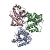
| ||||||||
|---|---|---|---|---|---|---|---|---|---|
| 1 | 
| ||||||||
| 2 | 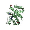
| ||||||||
| 3 | 
| ||||||||
| Unit cell |
|
-Components
| #1: Protein | Mass: 23801.539 Da / Num. of mol.: 3 Source method: isolated from a genetically manipulated source Source: (gene. exp.) Homo sapiens (human) / Gene: TRMT10C, MRPP1, RG9MTD1 / Plasmid: pNIC28-Bsa4 / Production host:  Escherichia coli (E. coli) Escherichia coli (E. coli)References: UniProt: Q7L0Y3, Transferases; Transferring one-carbon groups; Methyltransferases#2: Chemical | S-Adenosyl methionine  Mass: 398.437 Da / Num. of mol.: 3 / Source method: obtained synthetically / Formula: C15H22N6O5S Mass: 398.437 Da / Num. of mol.: 3 / Source method: obtained synthetically / Formula: C15H22N6O5S#3: Chemical | ChemComp-GOL / Glycerol  Mass: 92.094 Da / Num. of mol.: 7 / Source method: obtained synthetically / Formula: C3H8O3 Mass: 92.094 Da / Num. of mol.: 7 / Source method: obtained synthetically / Formula: C3H8O3#4: Chemical | ChemComp-EDO / Ethylene glycol  Mass: 62.068 Da / Num. of mol.: 4 / Source method: obtained synthetically / Formula: C2H6O2 Mass: 62.068 Da / Num. of mol.: 4 / Source method: obtained synthetically / Formula: C2H6O2#5: Water | ChemComp-HOH / | Water Mass: 18.015 Da / Num. of mol.: 217 / Source method: isolated from a natural source / Formula: H2O Mass: 18.015 Da / Num. of mol.: 217 / Source method: isolated from a natural source / Formula: H2O |
|---|
-Experimental details
-Experiment
| Experiment | Method: X-RAY DIFFRACTION / Number of used crystals: 1 |
|---|
- Sample preparation
| Crystal | Density Matthews: 3.43 Å3/Da / Density % sol: 64.2 % / Description: Diamonds |
|---|---|
| Crystal grow | Temperature: 277.15 K / Method: vapor diffusion, sitting drop / pH: 7.5 Details: Well solution: 12% PEG 1000, 28% glycerol, 1.5%(w/v) PEG 3350 Cryo: 25% propylene glycol Protein concentration: 11 mg/mL |
-Data collection
| Diffraction | Mean temperature: 100 K |
|---|---|
| Diffraction source | Source: SYNCHROTRON / Site: Diamond  / Beamline: I04-1 / Wavelength: 0.9282 Å / Beamline: I04-1 / Wavelength: 0.9282 Å |
| Detector | Type: DECTRIS PILATUS 6M-F / Detector: PIXEL / Date: Oct 23, 2015 |
| Radiation | Protocol: SINGLE WAVELENGTH / Monochromatic (M) / Laue (L): M / Scattering type: x-ray |
| Radiation wavelength | Wavelength: 0.9282 Å / Relative weight: 1 |
| Reflection | Resolution: 1.96→82.64 Å / Num. obs: 67692 / % possible obs: 99.98 % / Redundancy: 6.9 % / CC1/2: 0.999 / Rmerge(I) obs: 0.052 / Net I/σ(I): 16.95 |
- Processing
| Software |
| ||||||||||||||||||||||||||||||||||||||||||||||||||||||||||||||||||||||||||||||||||||||||||||||||||||||||||||||||||||||||||||||||||||||||||||||||||||||||||||||||||||||||||||||||||||||
|---|---|---|---|---|---|---|---|---|---|---|---|---|---|---|---|---|---|---|---|---|---|---|---|---|---|---|---|---|---|---|---|---|---|---|---|---|---|---|---|---|---|---|---|---|---|---|---|---|---|---|---|---|---|---|---|---|---|---|---|---|---|---|---|---|---|---|---|---|---|---|---|---|---|---|---|---|---|---|---|---|---|---|---|---|---|---|---|---|---|---|---|---|---|---|---|---|---|---|---|---|---|---|---|---|---|---|---|---|---|---|---|---|---|---|---|---|---|---|---|---|---|---|---|---|---|---|---|---|---|---|---|---|---|---|---|---|---|---|---|---|---|---|---|---|---|---|---|---|---|---|---|---|---|---|---|---|---|---|---|---|---|---|---|---|---|---|---|---|---|---|---|---|---|---|---|---|---|---|---|---|---|---|---|
| Refinement | Method to determine structure: SAD / Resolution: 1.96→82.64 Å / Cor.coef. Fo:Fc: 0.971 / Cor.coef. Fo:Fc free: 0.964 / SU B: 4.148 / SU ML: 0.108 / Cross valid method: THROUGHOUT / ESU R: 0.116 / ESU R Free: 0.109 / Details: HYDROGENS HAVE BEEN ADDED IN THE RIDING POSITIONS
| ||||||||||||||||||||||||||||||||||||||||||||||||||||||||||||||||||||||||||||||||||||||||||||||||||||||||||||||||||||||||||||||||||||||||||||||||||||||||||||||||||||||||||||||||||||||
| Solvent computation | Ion probe radii: 0.8 Å / Shrinkage radii: 0.8 Å / VDW probe radii: 1.2 Å | ||||||||||||||||||||||||||||||||||||||||||||||||||||||||||||||||||||||||||||||||||||||||||||||||||||||||||||||||||||||||||||||||||||||||||||||||||||||||||||||||||||||||||||||||||||||
| Displacement parameters | Biso mean: 53.966 Å2
| ||||||||||||||||||||||||||||||||||||||||||||||||||||||||||||||||||||||||||||||||||||||||||||||||||||||||||||||||||||||||||||||||||||||||||||||||||||||||||||||||||||||||||||||||||||||
| Refinement step | Cycle: 1 / Resolution: 1.96→82.64 Å
| ||||||||||||||||||||||||||||||||||||||||||||||||||||||||||||||||||||||||||||||||||||||||||||||||||||||||||||||||||||||||||||||||||||||||||||||||||||||||||||||||||||||||||||||||||||||
| Refine LS restraints |
|