 Movie
Movie Controller

[English] 日本語
 Yorodumi
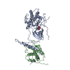
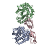
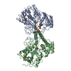
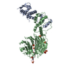
- PDB-5ncl: Crystal structure of the Cbk1-Mob2 kinase-coactivator complex wit... -
+ Open data

- Basic information
| Entry | Database: PDB / ID: 5ncl | ||||||
|---|---|---|---|---|---|---|---|
| Title | Crystal structure of the Cbk1-Mob2 kinase-coactivator complex with an SSD1 peptide | ||||||
 Components Components |
| ||||||
 Keywords Keywords |  SIGNALING PROTEIN / kinase SIGNALING PROTEIN / kinase | ||||||
| Function / homology |  Function and homology information Function and homology informationregulation of fungal-type cell wall organization / budding cell apical bud growth / cellular bud / prospore membrane / establishment or maintenance of actin cytoskeleton polarity / : / exosome (RNase complex) / incipient cellular bud site / septum digestion after cytokinesis / intracellular mRNA localization ...regulation of fungal-type cell wall organization / budding cell apical bud growth / cellular bud / prospore membrane / establishment or maintenance of actin cytoskeleton polarity / : / exosome (RNase complex) / incipient cellular bud site / septum digestion after cytokinesis / intracellular mRNA localization / cellular bud tip / serine/threonine protein kinase complex / : / cellular bud neck / regulation of G1 to G0 transition / mating projection tip / establishment or maintenance of cell polarity / protein kinase activator activity / regulation of protein secretion / mRNA catabolic process / mRNA regulatory element binding translation repressor activity / mRNA 3'-UTR binding / P-body / mRNA 5'-UTR binding / cytoplasmic stress granule / cell cortex / 3'-5'-RNA exonuclease activity / negative regulation of translation / non-specific serine/threonine protein kinase / intracellular signal transduction / positive regulation of protein phosphorylation / cell cycle / cell division / phosphorylation / protein serine kinase activity / mRNA binding / protein serine/threonine kinase activity / signal transduction / ATP binding / identical protein binding / nucleus / cytoplasmSimilarity search - Function | ||||||
| Biological species |  Saccharomyces cerevisiae (brewer's yeast) Saccharomyces cerevisiae (brewer's yeast) | ||||||
| Method | X-RAY DIFFRACTION / SYNCHROTRON / MOLECULAR REPLACEMENT / Resolution: 3.15 Å | ||||||
 Authors Authors | Gogl, G. / Remenyi, A. / Parker, B. / Weiss, E. | ||||||
 Citation Citation | Journal: Biochemistry / Year: 2020 Title: Ndr/Lats Kinases Bind Specific Mob-Family Coactivators through a Conserved and Modular Interface. Authors: Parker, B.W. / Gogl, G. / Balint, M. / Hetenyi, C. / Remenyi, A. / Weiss, E.L. | ||||||
| History |
|
- Structure visualization
| Structure viewer | Molecule: MolmilJmol/JSmol |
|---|
- Downloads & links
-Download
| PDBx/mmCIF format | 5ncl.cif.gz | 264.7 KB | Display | PDBx/mmCIF format |
|---|---|---|---|---|
| PDB format | pdb5ncl.ent.gz | 208.7 KB | Display | PDB format |
| PDBx/mmJSON format | 5ncl.json.gz | Tree view | PDBx/mmJSON format | |
| Others |  Other downloads Other downloads |
-Validation report
| Arichive directory | https://data.pdbj.org/pub/pdb/validation_reports/nc/5nclftp://data.pdbj.org/pub/pdb/validation_reports/nc/5ncl | HTTPS FTP |
|---|
-Related structure data
| Related structure data |  5ncmC  5ncnC 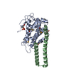 5brkS S: Starting model for refinement C: citing same article ( |
|---|---|
| Similar structure data |


-Links
 PDBj
PDBj


- Assembly
| Deposited unit | 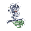
| ||||||||
|---|---|---|---|---|---|---|---|---|---|
| 1 |
| ||||||||
| Unit cell |
|
-Components
| #1: Protein | Mass: 58756.566 Da / Num. of mol.: 1 / Fragment: UNP Residues 251-756 Source method: isolated from a genetically manipulated source Source: (gene. exp.) Saccharomyces cerevisiae (brewer's yeast)Gene: CBK1, YNL161W, N1727 / Production host:  Escherichia coli (E. coli) Escherichia coli (E. coli)References: UniProt: P53894, non-specific serine/threonine protein kinase |
|---|---|
| #2: Protein | Mass: 28415.197 Da / Num. of mol.: 1 / Fragment: UNP Residues 46-287 Source method: isolated from a genetically manipulated source Source: (gene. exp.) Saccharomyces cerevisiae (brewer's yeast)Gene: MOB2, YFL034C-B, YFL035C, YFL035C-A / Production host: Escherichia coli (E. coli) / References: UniProt: P43563 |
| #3: Protein/peptide | Mass: 1200.273 Da / Num. of mol.: 1 / Fragment: UNP Residues 205-214 Source method: isolated from a genetically manipulated source Source: (gene. exp.) Saccharomyces cerevisiae (brewer's yeast)Gene: SSD1, CLA1, RLD1, SRK1, YDR293C, D9819.4 / Production host: Escherichia coli (E. coli) / References: UniProt: P24276 |
| #4: Chemical | ChemComp-ANP /   Mass: 506.196 Da / Num. of mol.: 1 / Source method: obtained synthetically / Formula: C10H17N6O12P3 / Comment: AMP-PNP, energy-carrying molecule analogue*YM Mass: 506.196 Da / Num. of mol.: 1 / Source method: obtained synthetically / Formula: C10H17N6O12P3 / Comment: AMP-PNP, energy-carrying molecule analogue*YM |
-Experimental details
-Experiment
| Experiment | Method: X-RAY DIFFRACTION / Number of used crystals: 1 |
|---|
- Sample preparation
| Crystal | Density Matthews: 4.06 Å3/Da / Density % sol: 69.7 % |
|---|---|
| Crystal grow | Temperature: 296 K / Method: microbatch / pH: 5.5 / Details: 25% PEG 20,000 BUFFERED WITH 0.1M NA-CITRATE |
-Data collection
| Diffraction | Mean temperature: 100 K |
|---|---|
| Diffraction source | Source: SYNCHROTRON / Site: SLS  / Beamline: X06DA / Wavelength: 1 Å / Beamline: X06DA / Wavelength: 1 Å |
| Detector | Type: DECTRIS PILATUS 2M / Detector: PIXEL / Date: Jun 30, 2012 |
| Radiation | Protocol: SINGLE WAVELENGTH / Monochromatic (M) / Laue (L): M / Scattering type: x-ray |
| Radiation wavelength | Wavelength: 1 Å / Relative weight: 1 |
| Reflection | Resolution: 3.15→44.595 Å / Num. obs: 19888 / % possible obs: 99.7 % / Redundancy: 7.7 % / CC1/2: 0.996 / Rmerge(I) obs: 0.168 / Net I/σ(I): 9.37 |
| Reflection shell | Resolution: 3.15→3.23 Å / Redundancy: 7.8 % / Rmerge(I) obs: 1.04 / Mean I/σ(I) obs: 2.05 / Num. unique obs: 1455 / CC1/2: 0.804 / % possible all: 99.7 |
- Processing
| Software |
| ||||||||||||||||||||||||||||||||||||||||||||||||||||||||
|---|---|---|---|---|---|---|---|---|---|---|---|---|---|---|---|---|---|---|---|---|---|---|---|---|---|---|---|---|---|---|---|---|---|---|---|---|---|---|---|---|---|---|---|---|---|---|---|---|---|---|---|---|---|---|---|---|---|
| Refinement | Method to determine structure: MOLECULAR REPLACEMENT Starting model: 5BRK Resolution: 3.15→44.595 Å / SU ML: 0.54 / Cross valid method: FREE R-VALUE / σ(F): 1.35 / Phase error: 35.74
| ||||||||||||||||||||||||||||||||||||||||||||||||||||||||
| Solvent computation | Shrinkage radii: 0.9 Å / VDW probe radii: 1.11 Å | ||||||||||||||||||||||||||||||||||||||||||||||||||||||||
| Refinement step | Cycle: LAST / Resolution: 3.15→44.595 Å
| ||||||||||||||||||||||||||||||||||||||||||||||||||||||||
| Refine LS restraints |
| ||||||||||||||||||||||||||||||||||||||||||||||||||||||||
| LS refinement shell |
| ||||||||||||||||||||||||||||||||||||||||||||||||||||||||
| Refinement TLS params. | Method: refined / Origin x: -20.7981 Å / Origin y: -13.6636 Å / Origin z: 19.783 Å
| ||||||||||||||||||||||||||||||||||||||||||||||||||||||||
| Refinement TLS group | Selection details: all |
