 Movie
Movie Controller

[English] 日本語
 Yorodumi
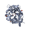
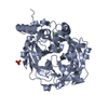
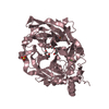
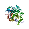
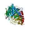
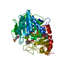
- PDB-6vhq: Crystal structure of Bacillus subtilis levansucrase (D86A/E342A) ... -
+ Open data

- Basic information
| Entry | Database: PDB / ID: 6vhq | ||||||||||||
|---|---|---|---|---|---|---|---|---|---|---|---|---|---|
| Title | Crystal structure of Bacillus subtilis levansucrase (D86A/E342A) in complex with oligosaccharides | ||||||||||||
 Components Components | Glycoside hydrolase family 68 protein | ||||||||||||
 Keywords Keywords |  TRANSFERASE / Levansucrase / Glycoside hydrolase / Levan / Fructose polymers TRANSFERASE / Levansucrase / Glycoside hydrolase / Levan / Fructose polymers | ||||||||||||
| Function / homology |  Function and homology informationlevansucrase / levansucrase activity / carbohydrate utilization / extracellular region Function and homology informationlevansucrase / levansucrase activity / carbohydrate utilization / extracellular regionSimilarity search - Function | ||||||||||||
| Biological species |  Bacillus subtilis (bacteria) Bacillus subtilis (bacteria) | ||||||||||||
| Method | X-RAY DIFFRACTION / MOLECULAR REPLACEMENT / Resolution: 2.047 Å | ||||||||||||
 Authors Authors | Diaz-Vilchis, A. / Raga-Carbajal, E. / Rojas-Trejo, S. / Olvera, C. / Rudino-Pinera, E. | ||||||||||||
| Funding support |  Mexico, 3items Mexico, 3items
| ||||||||||||
 Citation Citation | Journal: J.Biol.Chem. / Year: 2020 Title: The molecular basis of the nonprocessive elongation mechanism in levansucrases. Authors: Raga-Carbajal, E. / Diaz-Vilchis, A. / Rojas-Trejo, S.P. / Rudino-Pinera, E. / Olvera, C. | ||||||||||||
| History |
|
- Structure visualization
| Structure viewer | Molecule: MolmilJmol/JSmol |
|---|
- Downloads & links
-Download
| PDBx/mmCIF format | 6vhq.cif.gz | 209.3 KB | Display | PDBx/mmCIF format |
|---|---|---|---|---|
| PDB format | pdb6vhq.ent.gz | 165.4 KB | Display | PDB format |
| PDBx/mmJSON format | 6vhq.json.gz | Tree view | PDBx/mmJSON format | |
| Others |  Other downloads Other downloads |
-Validation report
| Arichive directory | https://data.pdbj.org/pub/pdb/validation_reports/vh/6vhqftp://data.pdbj.org/pub/pdb/validation_reports/vh/6vhq | HTTPS FTP |
|---|
-Related structure data
| Related structure data | 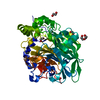 1oygS S: Starting model for refinement |
|---|---|
| Similar structure data |

-Links
 PDBj
PDBj

- Assembly
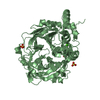
| Deposited unit | 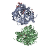
| ||||||||
|---|---|---|---|---|---|---|---|---|---|
| 1 | 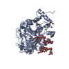
| ||||||||
| 2 | 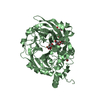
| ||||||||
| Unit cell |
|
-Components
-Protein , 1 types, 2 molecules AB
| #1: Protein | Mass: 52360.797 Da / Num. of mol.: 2 / Mutation: D86A, E342A Source method: isolated from a genetically manipulated source Source: (gene. exp.) Bacillus subtilis (bacteria)Gene: sacB, B4417_3269, ETA10_18085, ETL41_09350, FVD40_16900 Production host: Escherichia coli BL21 (bacteria) / References: UniProt: A0PFL2, UniProt: P05655*PLUS |
|---|
-Sugars , 2 types, 4 molecules
| #2: Polysaccharide | / Mass: 990.860 Da / Num. of mol.: 3 Source method: isolated from a genetically manipulated source #3: Polysaccharide | beta-D-fructofuranose-(2-6)-beta-D-fructofuranose / levanbiose | 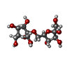  , Oligosaccharide / Class: Metabolism / Mass: 342.297 Da / Num. of mol.: 1 , Oligosaccharide / Class: Metabolism / Mass: 342.297 Da / Num. of mol.: 1Source method: isolated from a genetically manipulated source Details: oligosaccharide / References: levanbiose |
|---|
-Non-polymers , 3 types, 551 molecules 


| #4: Chemical |  Mass: 40.078 Da / Num. of mol.: 2 / Source method: obtained synthetically / Formula: Ca / Feature type: SUBJECT OF INVESTIGATION Mass: 40.078 Da / Num. of mol.: 2 / Source method: obtained synthetically / Formula: Ca / Feature type: SUBJECT OF INVESTIGATION#5: Chemical | ChemComp-BR / Bromide Mass: 79.904 Da / Num. of mol.: 9 / Source method: obtained synthetically / Formula: Br / Feature type: SUBJECT OF INVESTIGATION Mass: 79.904 Da / Num. of mol.: 9 / Source method: obtained synthetically / Formula: Br / Feature type: SUBJECT OF INVESTIGATION#6: Water | ChemComp-HOH / | WaterMass: 18.015 Da / Num. of mol.: 540 / Source method: isolated from a natural source / Formula: H2O |
|---|
-Details
| Has ligand of interest | Y |
|---|---|
| Nonpolymer details | The entry contains the oligosaccharide levanhexaose, a 6 fructose units bound by beta2-6 glycosidic bonds |
-Experimental details
-Experiment
| Experiment | Method: X-RAY DIFFRACTION / Number of used crystals: 1 |
|---|
- Sample preparation
| Crystal | Density Matthews: 2.17 Å3/Da / Density % sol: 43.22 % |
|---|---|
| Crystal grow | Temperature: 291 K / Method: microbatch / pH: 6 / Details: 30 % w/v PEG 2000 MME and 150 mM Potassium Bromide |
-Data collection
| Diffraction | Mean temperature: 100 K / Serial crystal experiment: N |
|---|---|
| Diffraction source | Source: ROTATING ANODE / Type: RIGAKU / Wavelength: 1.54 Å |
| Detector | Type: DECTRIS PILATUS 200K / Detector: PIXEL / Date: Aug 10, 2016 |
| Radiation | Protocol: SINGLE WAVELENGTH / Monochromatic (M) / Laue (L): M / Scattering type: x-ray |
| Radiation wavelength | Wavelength: 1.54 Å / Relative weight: 1 |
| Reflection | Resolution: 2.047→35.205 Å / Num. obs: 53175 / % possible obs: 99.99 % / Observed criterion σ(I): 3.5 / Redundancy: 2.7 % / Biso Wilson estimate: 25.1 Å2 / Rmerge(I) obs: 0.048 / Net I/σ(I): 23.9 |
| Reflection shell | Resolution: 2.047→2.09 Å / Redundancy: 2.5 % / Rmerge(I) obs: 0.526 / Mean I/σ(I) obs: 3.5 / Num. unique obs: 5145 / % possible all: 100 |
- Processing
| Software |
| ||||||||||||||||||||||||||||||||||||||||||||||||||||||||||||||||||||||||||||||||||||||||||||||||||||||||||||||||||||||||
|---|---|---|---|---|---|---|---|---|---|---|---|---|---|---|---|---|---|---|---|---|---|---|---|---|---|---|---|---|---|---|---|---|---|---|---|---|---|---|---|---|---|---|---|---|---|---|---|---|---|---|---|---|---|---|---|---|---|---|---|---|---|---|---|---|---|---|---|---|---|---|---|---|---|---|---|---|---|---|---|---|---|---|---|---|---|---|---|---|---|---|---|---|---|---|---|---|---|---|---|---|---|---|---|---|---|---|---|---|---|---|---|---|---|---|---|---|---|---|---|---|---|
| Refinement | Method to determine structure: MOLECULAR REPLACEMENT Starting model: 1OYG Resolution: 2.047→35.205 Å / SU ML: 0.22 / Cross valid method: FREE R-VALUE / σ(F): 1.35 / Phase error: 24.23
| ||||||||||||||||||||||||||||||||||||||||||||||||||||||||||||||||||||||||||||||||||||||||||||||||||||||||||||||||||||||||
| Solvent computation | Shrinkage radii: 0.9 Å / VDW probe radii: 1.11 Å | ||||||||||||||||||||||||||||||||||||||||||||||||||||||||||||||||||||||||||||||||||||||||||||||||||||||||||||||||||||||||
| Displacement parameters | Biso max: 94.72 Å2 / Biso mean: 28.2683 Å2 / Biso min: 7.12 Å2 | ||||||||||||||||||||||||||||||||||||||||||||||||||||||||||||||||||||||||||||||||||||||||||||||||||||||||||||||||||||||||
| Refinement step | Cycle: final / Resolution: 2.047→35.205 Å
| ||||||||||||||||||||||||||||||||||||||||||||||||||||||||||||||||||||||||||||||||||||||||||||||||||||||||||||||||||||||||
| Refine LS restraints |
| ||||||||||||||||||||||||||||||||||||||||||||||||||||||||||||||||||||||||||||||||||||||||||||||||||||||||||||||||||||||||
| LS refinement shell | Refine-ID: X-RAY DIFFRACTION / Rfactor Rfree error: 0
|
