 Movie
Movie Controller


+ Open data

- Basic information
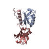
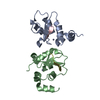
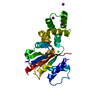
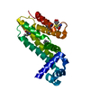
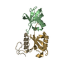
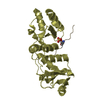
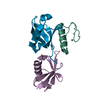
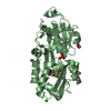
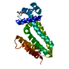
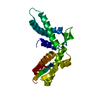
| Entry | Database: PDB / ID: 5n88 | ||||||
|---|---|---|---|---|---|---|---|
| Title | Crystal structure of antibody bound to viral protein | ||||||
 Components Components |
| ||||||
 Keywords Keywords |  IMMUNE SYSTEM / MLL: HIV: intracellular antibody: integrase: LEDGF / AIDS IMMUNE SYSTEM / MLL: HIV: intracellular antibody: integrase: LEDGF / AIDS | ||||||
| Function / homology |  Function and homology informationsupercoiled DNA binding / Integration of viral DNA into host genomic DNA / Autointegration results in viral DNA circles / Formation of WDR5-containing histone-modifying complexes / 2-LTR circle formation / Vpr-mediated nuclear import of PICs / Integration of provirus / APOBEC3G mediated resistance to HIV-1 infection / mRNA 5'-splice site recognition / heterochromatin ...supercoiled DNA binding / Integration of viral DNA into host genomic DNA / Autointegration results in viral DNA circles / Formation of WDR5-containing histone-modifying complexes / 2-LTR circle formation / Vpr-mediated nuclear import of PICs / Integration of provirus / APOBEC3G mediated resistance to HIV-1 infection / mRNA 5'-splice site recognition / heterochromatin / nuclear periphery / euchromatin / response to heat / DNA-binding transcription factor binding / response to oxidative stress / transcription coactivator activity / chromatin remodeling / chromatin binding / positive regulation of transcription by RNA polymerase II / RNA binding / nucleoplasm / nucleus / cytosol Function and homology informationsupercoiled DNA binding / Integration of viral DNA into host genomic DNA / Autointegration results in viral DNA circles / Formation of WDR5-containing histone-modifying complexes / 2-LTR circle formation / Vpr-mediated nuclear import of PICs / Integration of provirus / APOBEC3G mediated resistance to HIV-1 infection / mRNA 5'-splice site recognition / heterochromatin ...supercoiled DNA binding / Integration of viral DNA into host genomic DNA / Autointegration results in viral DNA circles / Formation of WDR5-containing histone-modifying complexes / 2-LTR circle formation / Vpr-mediated nuclear import of PICs / Integration of provirus / APOBEC3G mediated resistance to HIV-1 infection / mRNA 5'-splice site recognition / heterochromatin / nuclear periphery / euchromatin / response to heat / DNA-binding transcription factor binding / response to oxidative stress / transcription coactivator activity / chromatin remodeling / chromatin binding / positive regulation of transcription by RNA polymerase II / RNA binding / nucleoplasm / nucleus / cytosolSimilarity search - Function | ||||||
| Biological species |  Homo sapiens (human) Homo sapiens (human) | ||||||
| Method | X-RAY DIFFRACTION / SYNCHROTRON / MOLECULAR REPLACEMENT / molecular replacement / Resolution: 1.7 Å | ||||||
 Authors Authors | Bao, L. / Hannon, C. / Cruz-Migoni, A. / Ptchelkine, D. / Sun, M.-y. / Derveni, M. / Bunjobpol, W. / Chambers, J.S. / Simmons, A. / Phillips, S.E.V. / Rabbitts, T.H. | ||||||
 Citation Citation | Journal: Sci Rep / Year: 2017 Title: Intracellular immunization against HIV infection with an intracellular antibody that mimics HIV integrase binding to the cellular LEDGF protein. Authors: Bao, L. / Hannon, C. / Cruz-Mignoni, A. / Ptchelkine, D. / Sun, M.Y. / Miller, A. / Bunjobpol, W. / Quevedo, C.E. / Derveni, M. / Chambers, J. / Simmons, A. / Phillips, S.E.V. / Rabbitts, T.H. | ||||||
| History |
|
- Structure visualization
| Structure viewer | Molecule: MolmilJmol/JSmol |
|---|
- Downloads & links
-Download
| PDBx/mmCIF format | 5n88.cif.gz | 96.9 KB | Display | PDBx/mmCIF format |
|---|---|---|---|---|
| PDB format | pdb5n88.ent.gz | 77.5 KB | Display | PDB format |
| PDBx/mmJSON format | 5n88.json.gz | Tree view | PDBx/mmJSON format | |
| Others |  Other downloads Other downloads |
-Validation report
| Arichive directory | https://data.pdbj.org/pub/pdb/validation_reports/n8/5n88ftp://data.pdbj.org/pub/pdb/validation_reports/n8/5n88 | HTTPS FTP |
|---|
-Related structure data
| Similar structure data |
|---|
-Links
 PDBj
PDBj


- Assembly
| Deposited unit | 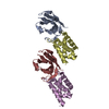
| ||||||||
|---|---|---|---|---|---|---|---|---|---|
| 1 | 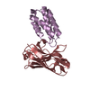
| ||||||||
| 2 | 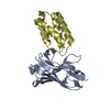
| ||||||||
| Unit cell |
|
-Components
| #1: Antibody | Mass: 13886.484 Da / Num. of mol.: 2 Source method: isolated from a genetically manipulated source Source: (gene. exp.) Homo sapiens (human) / Gene: PSIP1 / Plasmid: pRK-172 / Production host:  Escherichia coli BL21(DE3) (bacteria) / Variant (production host): C41 Escherichia coli BL21(DE3) (bacteria) / Variant (production host): C41#2: Protein | | Mass: 9310.934 Da / Num. of mol.: 1 Source method: isolated from a genetically manipulated source Source: (gene. exp.) Homo sapiens (human) / Gene: PSIP1, DFS70, LEDGF, PSIP2 / Plasmid: pRK-172 / Production host: Escherichia coli BL21(DE3) (bacteria) / Variant (production host): C41 / References: UniProt: O75475#3: Protein | | Mass: 9340.961 Da / Num. of mol.: 1 Source method: isolated from a genetically manipulated source Source: (gene. exp.) Homo sapiens (human) / Gene: PSIP1, DFS70, LEDGF, PSIP2 / Plasmid: pRK-172 / Production host: Escherichia coli BL21(DE3) (bacteria) / Variant (production host): C41 / References: UniProt: O75475#4: Water | ChemComp-HOH / | Water Mass: 18.015 Da / Num. of mol.: 320 / Source method: isolated from a natural source / Formula: H2O Mass: 18.015 Da / Num. of mol.: 320 / Source method: isolated from a natural source / Formula: H2O |
|---|
-Experimental details
-Experiment
| Experiment | Method: X-RAY DIFFRACTION / Number of used crystals: 1 |
|---|
- Sample preparation
| Crystal | Density Matthews: 1.73 Å3/Da / Density % sol: 28.83 % |
|---|---|
| Crystal grow | Temperature: 277 K / Method: vapor diffusion / pH: 8.5 / Details: 25.0% PEG 3350, 10mM Tris pH8.5 |
-Data collection
| Diffraction | Mean temperature: 100 K |
|---|---|
| Diffraction source | Source: SYNCHROTRON / Site: Diamond  / Beamline: I04-1 / Wavelength: 0.9795 Å / Beamline: I04-1 / Wavelength: 0.9795 Å |
| Detector | Type: DECTRIS PILATUS 6M-F / Detector: PIXEL / Date: Apr 29, 2013 |
| Radiation | Protocol: SINGLE WAVELENGTH / Monochromatic (M) / Laue (L): M / Scattering type: x-ray |
| Radiation wavelength | Wavelength: 0.9795 Å / Relative weight: 1 |
| Reflection | Resolution: 1.7→15 Å / Num. obs: 115544 / % possible obs: 90.4 % / Redundancy: 3.73 % / Net I/σ(I): 2.9 |
-Phasing
| Phasing | Method: molecular replacement |
|---|
- Processing
| Software |
| ||||||||||||||||||||||||||||||||||||||||||||||||||||||||||||
|---|---|---|---|---|---|---|---|---|---|---|---|---|---|---|---|---|---|---|---|---|---|---|---|---|---|---|---|---|---|---|---|---|---|---|---|---|---|---|---|---|---|---|---|---|---|---|---|---|---|---|---|---|---|---|---|---|---|---|---|---|---|
| Refinement | Method to determine structure: MOLECULAR REPLACEMENT / Resolution: 1.7→14.92 Å / Cor.coef. Fo:Fc: 0.95 / Cor.coef. Fo:Fc free: 0.911 / SU B: 3.29 / SU ML: 0.108 / Cross valid method: THROUGHOUT / σ(F): 0 / ESU R: 0.169 / ESU R Free: 0.16 / Details: Refmac5
| ||||||||||||||||||||||||||||||||||||||||||||||||||||||||||||
| Solvent computation | Ion probe radii: 0.8 Å / Shrinkage radii: 0.8 Å / VDW probe radii: 1.2 Å | ||||||||||||||||||||||||||||||||||||||||||||||||||||||||||||
| Displacement parameters | Biso max: 76.12 Å2 / Biso mean: 20.012 Å2 / Biso min: 7.19 Å2
| ||||||||||||||||||||||||||||||||||||||||||||||||||||||||||||
| Refinement step | Cycle: final / Resolution: 1.7→14.92 Å
| ||||||||||||||||||||||||||||||||||||||||||||||||||||||||||||
| Refine LS restraints |
| ||||||||||||||||||||||||||||||||||||||||||||||||||||||||||||
| LS refinement shell | Resolution: 1.7→1.744 Å / Rfactor Rfree error: 0 / Total num. of bins used: 20
|
