 Movie
Movie Controller


+ Open data

- Basic information
| Entry | Database: PDB / ID: 5n6f | |||||||||
|---|---|---|---|---|---|---|---|---|---|---|
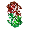
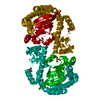
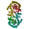
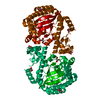
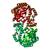
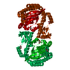
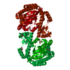
| Title | Crystal structure of TGT in complex with guanine fragment | |||||||||
 Components Components | Queuine tRNA-ribosyltransferase | |||||||||
 Keywords Keywords | TRANSFERASE / TGT / TRNA / GUANINE EXCHANGE ENZYME / TRANSGLYCOSYLASE | |||||||||
| Function / homology |  Function and homology information Function and homology informationtRNA-guanosine34 preQ1 transglycosylase / tRNA wobble guanine modification / tRNA-guanosine(34) queuine transglycosylase activity / tRNA-guanine transglycosylation / queuosine biosynthetic process / metal ion binding / cytosolSimilarity search - Function | |||||||||
| Biological species |  Zymomonas mobilis subsp. mobilis (bacteria) Zymomonas mobilis subsp. mobilis (bacteria) | |||||||||
| Method | X-RAY DIFFRACTION / SYNCHROTRON / MOLECULAR REPLACEMENT / Resolution: 1.11909520042 Å | |||||||||
 Authors Authors | Hassaan, E. / Heine, A. / Klebe, G. | |||||||||
 Citation Citation | Journal: Chemmedchem / Year: 2020 Title: Fragments as Novel Starting Points for tRNA-Guanine Transglycosylase Inhibitors Found by Alternative Screening Strategies. Authors: Hassaan, E. / Eriksson, P.O. / Geschwindner, S. / Heine, A. / Klebe, G. | |||||||||
| History |
|
- Structure visualization
| Structure viewer | Molecule: MolmilJmol/JSmol |
|---|
- Downloads & links
-Download
| PDBx/mmCIF format | 5n6f.cif.gz | 277.1 KB | Display | PDBx/mmCIF format |
|---|---|---|---|---|
| PDB format | pdb5n6f.ent.gz | 185.2 KB | Display | PDB format |
| PDBx/mmJSON format | 5n6f.json.gz | Tree view | PDBx/mmJSON format | |
| Others |  Other downloads Other downloads |
-Validation report
| Arichive directory | https://data.pdbj.org/pub/pdb/validation_reports/n6/5n6fftp://data.pdbj.org/pub/pdb/validation_reports/n6/5n6f | HTTPS FTP |
|---|
-Related structure data
| Related structure data | 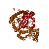 5sw3C 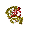 5utiC 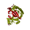 5utjC 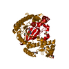 5v3cC 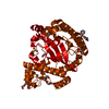 6fsoC 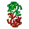 4lbuS S: Starting model for refinement C: citing same article ( |
|---|---|
| Similar structure data |


-Links
 PDBj
PDBj

- Assembly
| Deposited unit | 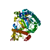
| ||||||||||
|---|---|---|---|---|---|---|---|---|---|---|---|
| 1 | 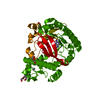
| ||||||||||
| Unit cell |
|
-Components
-Protein , 1 types, 1 molecules A
| #1: Protein | / Guanine insertion enzyme / tRNA-guanine transglycosylase Mass: 41763.484 Da / Num. of mol.: 1 Source method: isolated from a genetically manipulated source Source: (gene. exp.) Zymomonas mobilis subsp. mobilis (strain ATCC 31821 / ZM4 / CP4) (bacteria)Strain: ATCC 31821 / ZM4 / CP4 / Gene: tgt, ZMO0363 / Plasmid: PPR-IBA2 / Production host: Escherichia coli BL21(DE3) (bacteria) / Variant (production host): CODONPLUSReferences: UniProt: P28720, tRNA-guanosine34 preQ1 transglycosylase |
|---|
-Non-polymers , 6 types, 284 molecules 


| #2: Chemical | ChemComp-ZN /  Mass: 65.409 Da / Num. of mol.: 1 / Source method: obtained synthetically / Formula: Zn Mass: 65.409 Da / Num. of mol.: 1 / Source method: obtained synthetically / Formula: Zn |
|---|---|
| #3: Chemical | ChemComp-DMS / Dimethyl sulfoxide Mass: 78.133 Da / Num. of mol.: 1 / Source method: obtained synthetically / Formula: C2H6OS / Comment: DMSO, precipitant*YM Mass: 78.133 Da / Num. of mol.: 1 / Source method: obtained synthetically / Formula: C2H6OS / Comment: DMSO, precipitant*YM |
| #4: Chemical | ChemComp-PEG / Diethylene glycol Mass: 106.120 Da / Num. of mol.: 1 / Source method: obtained synthetically / Formula: C4H10O3 Mass: 106.120 Da / Num. of mol.: 1 / Source method: obtained synthetically / Formula: C4H10O3 |
| #5: Chemical | ChemComp-GUN / Guanine Mass: 151.126 Da / Num. of mol.: 1 / Source method: obtained synthetically / Formula: C5H5N5O Mass: 151.126 Da / Num. of mol.: 1 / Source method: obtained synthetically / Formula: C5H5N5O |
| #6: Chemical | ChemComp-PGE / Polyethylene glycol Mass: 150.173 Da / Num. of mol.: 1 / Source method: obtained synthetically / Formula: C6H14O4 Mass: 150.173 Da / Num. of mol.: 1 / Source method: obtained synthetically / Formula: C6H14O4 |
| #7: Water | ChemComp-HOH / WaterMass: 18.015 Da / Num. of mol.: 279 / Source method: isolated from a natural source / Formula: H2O |
-Details
| Has ligand of interest | Y |
|---|
-Experimental details
-Experiment
| Experiment | Method: X-RAY DIFFRACTION / Number of used crystals: 1 |
|---|
- Sample preparation
| Crystal | Density Matthews: 2.43 Å3/Da / Density % sol: 49.36 % |
|---|---|
| Crystal grow | Temperature: 289 K / Method: vapor diffusion, sitting drop / pH: 5.5 / Details: 13% PEG8000, 100MM MES, 1MM DTT, 10% DMSO |
-Data collection
| Diffraction | Mean temperature: 100 K / Serial crystal experiment: N |
|---|---|
| Diffraction source | Source: SYNCHROTRON / Site: BESSY  / Beamline: 14.1 / Wavelength: 0.918 Å / Beamline: 14.1 / Wavelength: 0.918 Å |
| Detector | Type: DECTRIS PILATUS 6M / Detector: PIXEL / Date: Jan 27, 2016 |
| Radiation | Protocol: SINGLE WAVELENGTH / Monochromatic (M) / Laue (L): M / Scattering type: x-ray |
| Radiation wavelength | Wavelength: 0.918 Å / Relative weight: 1 |
| Reflection | Resolution: 1.119→44.28 Å / Num. obs: 149008 / % possible obs: 97.1 % / Redundancy: 3.6 % / Biso Wilson estimate: 10.8545945983 Å2 / Rsym value: 0.061 / Net I/σ(I): 10.3 |
| Reflection shell | Resolution: 1.119→1.19 Å / Redundancy: 2.9 % / Rmerge(I) obs: 0.487 / Mean I/σ(I) obs: 1.96 / Num. unique obs: 149008 / % possible all: 83.2 |
- Processing
| Software |
| |||||||||||||||||||||||||||||||||||||||||||||||||||||||||||||||||||||||||||||||||||||||||||||||||||||||||||||||||||||||||||||||||||||||||||||||||||||||||||||||||||||||||||||||||||||||||||||||||||||||||||||||||||||||||
|---|---|---|---|---|---|---|---|---|---|---|---|---|---|---|---|---|---|---|---|---|---|---|---|---|---|---|---|---|---|---|---|---|---|---|---|---|---|---|---|---|---|---|---|---|---|---|---|---|---|---|---|---|---|---|---|---|---|---|---|---|---|---|---|---|---|---|---|---|---|---|---|---|---|---|---|---|---|---|---|---|---|---|---|---|---|---|---|---|---|---|---|---|---|---|---|---|---|---|---|---|---|---|---|---|---|---|---|---|---|---|---|---|---|---|---|---|---|---|---|---|---|---|---|---|---|---|---|---|---|---|---|---|---|---|---|---|---|---|---|---|---|---|---|---|---|---|---|---|---|---|---|---|---|---|---|---|---|---|---|---|---|---|---|---|---|---|---|---|---|---|---|---|---|---|---|---|---|---|---|---|---|---|---|---|---|---|---|---|---|---|---|---|---|---|---|---|---|---|---|---|---|---|---|---|---|---|---|---|---|---|---|---|---|---|---|---|---|---|
| Refinement | Method to determine structure: MOLECULAR REPLACEMENT Starting model: 4LBU Resolution: 1.11909520042→44.2793884339 Å / SU ML: 0.0973131262279 / Cross valid method: FREE R-VALUE / σ(F): 1.3545538695 / Phase error: 13.5056770047 Stereochemistry target values: GeoStd + Monomer Library + CDL v1.2
| |||||||||||||||||||||||||||||||||||||||||||||||||||||||||||||||||||||||||||||||||||||||||||||||||||||||||||||||||||||||||||||||||||||||||||||||||||||||||||||||||||||||||||||||||||||||||||||||||||||||||||||||||||||||||
| Solvent computation | Shrinkage radii: 0.9 Å / VDW probe radii: 1.11 Å / Solvent model: FLAT BULK SOLVENT MODEL | |||||||||||||||||||||||||||||||||||||||||||||||||||||||||||||||||||||||||||||||||||||||||||||||||||||||||||||||||||||||||||||||||||||||||||||||||||||||||||||||||||||||||||||||||||||||||||||||||||||||||||||||||||||||||
| Displacement parameters | Biso mean: 16.8179302453 Å2 | |||||||||||||||||||||||||||||||||||||||||||||||||||||||||||||||||||||||||||||||||||||||||||||||||||||||||||||||||||||||||||||||||||||||||||||||||||||||||||||||||||||||||||||||||||||||||||||||||||||||||||||||||||||||||
| Refinement step | Cycle: LAST / Resolution: 1.11909520042→44.2793884339 Å
| |||||||||||||||||||||||||||||||||||||||||||||||||||||||||||||||||||||||||||||||||||||||||||||||||||||||||||||||||||||||||||||||||||||||||||||||||||||||||||||||||||||||||||||||||||||||||||||||||||||||||||||||||||||||||
| Refine LS restraints |
| |||||||||||||||||||||||||||||||||||||||||||||||||||||||||||||||||||||||||||||||||||||||||||||||||||||||||||||||||||||||||||||||||||||||||||||||||||||||||||||||||||||||||||||||||||||||||||||||||||||||||||||||||||||||||
| LS refinement shell |
|
