 Movie
Movie Controller


+ Open data

- Basic information
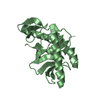
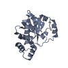
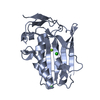
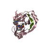
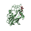
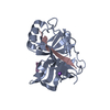
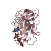
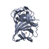
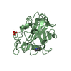
| Entry | Database: PDB / ID: 5mll | ||||||
|---|---|---|---|---|---|---|---|
| Title | Structure of HpDprA at 1.9 Angstroms resolution | ||||||
 Components Components | DNA processing chain A (DprA) | ||||||
 Keywords Keywords |  DNA BINDING PROTEIN / HpDprA DNA BINDING PROTEIN / HpDprA | ||||||
| Function / homology | DNA recombination-mediator protein A / DNA recombination-mediator protein A / DNA-mediated transformation / Rossmann fold - #450 / Rossmann fold / 3-Layer(aba) Sandwich / Alpha Beta / DNA processing chain A (DprA) Function and homology information Function and homology information | ||||||
| Biological species |  Helicobacter pylori 26695 (bacteria) Helicobacter pylori 26695 (bacteria) | ||||||
| Method | X-RAY DIFFRACTION / SYNCHROTRON / MOLECULAR REPLACEMENT / Resolution: 1.9 Å | ||||||
 Authors Authors | Lisboa, J. / Quevillon-Cheruel, S. | ||||||
 Citation Citation | Journal: FEBS J. / Year: 2019 Title: The C-terminal domain of HpDprA is a DNA-binding Winged Helix domain that does not bind double-stranded DNA. Authors: Lisboa, J. / Celma, L. / Sanchez, D. / Marquis, M. / Andreani, J. / Guerois, R. / Ochsenbein, F. / Durand, D. / Marsin, S. / Cuniasse, P. / Radicella, J.P. / Quevillon-Cheruel, S. | ||||||
| History |
|
- Structure visualization
| Structure viewer | Molecule: MolmilJmol/JSmol |
|---|
- Downloads & links
-Download
| PDBx/mmCIF format | 5mll.cif.gz | 99.9 KB | Display | PDBx/mmCIF format |
|---|---|---|---|---|
| PDB format | pdb5mll.ent.gz | 76.3 KB | Display | PDB format |
| PDBx/mmJSON format | 5mll.json.gz | Tree view | PDBx/mmJSON format | |
| Others |  Other downloads Other downloads |
-Validation report
| Arichive directory | https://data.pdbj.org/pub/pdb/validation_reports/ml/5mllftp://data.pdbj.org/pub/pdb/validation_reports/ml/5mll | HTTPS FTP |
|---|
-Related structure data
| Related structure data |  6gw7C 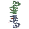 3uqzS S: Starting model for refinement C: citing same article ( |
|---|---|
| Similar structure data |

-Links
 PDBj
PDBj- Assembly
| Deposited unit | 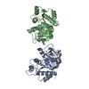
| ||||||||
|---|---|---|---|---|---|---|---|---|---|
| 1 | 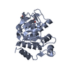
| ||||||||
| 2 | 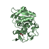
| ||||||||
| Unit cell |
|
-Components
| #1: Protein | Mass: 24450.414 Da / Num. of mol.: 2 Source method: isolated from a genetically manipulated source Source: (gene. exp.) Helicobacter pylori 26695 (bacteria) / Gene: HP_0333 / Production host: Escherichia coli (E. coli) / References: UniProt: O25100#2: Chemical | Ethylene glycol  Mass: 62.068 Da / Num. of mol.: 3 / Source method: obtained synthetically / Formula: C2H6O2 Mass: 62.068 Da / Num. of mol.: 3 / Source method: obtained synthetically / Formula: C2H6O2#3: Water | ChemComp-HOH / | Water Mass: 18.015 Da / Num. of mol.: 144 / Source method: isolated from a natural source / Formula: H2O Mass: 18.015 Da / Num. of mol.: 144 / Source method: isolated from a natural source / Formula: H2O |
|---|
-Experimental details
-Experiment
| Experiment | Method: X-RAY DIFFRACTION / Number of used crystals: 1 |
|---|
- Sample preparation
| Crystal | Density Matthews: 2.38 Å3/Da / Density % sol: 48.38 % |
|---|---|
| Crystal grow | Temperature: 293 K / Method: vapor diffusion, sitting drop / Details: 20% PEG 3350, 200mM di-ammonium tartrate |
-Data collection
| Diffraction | Mean temperature: 100 K |
|---|---|
| Diffraction source | Source: SYNCHROTRON / Site: SOLEIL  / Beamline: PROXIMA 1 / Wavelength: 0.98011 Å / Beamline: PROXIMA 1 / Wavelength: 0.98011 Å |
| Detector | Type: PSI PILATUS 6M / Detector: PIXEL / Date: Sep 30, 2011 |
| Radiation | Protocol: SINGLE WAVELENGTH / Monochromatic (M) / Laue (L): M / Scattering type: x-ray |
| Radiation wavelength | Wavelength: 0.98011 Å / Relative weight: 1 |
| Reflection | Resolution: 1.9→50 Å / Num. obs: 35234 / % possible obs: 99.4 % / Redundancy: 4.35 % / Biso Wilson estimate: 29.3 Å2 / Net I/σ(I): 3.2 |
| Reflection shell | Resolution: 1.9→1.966 Å / Redundancy: 4.35 % / % possible all: 96.98 |
- Processing
| Software |
| ||||||||||||||||||||||||||||||||||||||||||||||||||||||||||||||||||||||||||||||||||||||||||||||||||
|---|---|---|---|---|---|---|---|---|---|---|---|---|---|---|---|---|---|---|---|---|---|---|---|---|---|---|---|---|---|---|---|---|---|---|---|---|---|---|---|---|---|---|---|---|---|---|---|---|---|---|---|---|---|---|---|---|---|---|---|---|---|---|---|---|---|---|---|---|---|---|---|---|---|---|---|---|---|---|---|---|---|---|---|---|---|---|---|---|---|---|---|---|---|---|---|---|---|---|---|
| Refinement | Method to determine structure: MOLECULAR REPLACEMENT Starting model: 3UQZ Resolution: 1.9→41.753 Å / SU ML: 0.25 / Cross valid method: FREE R-VALUE / σ(F): 1.36 / Phase error: 26.05
| ||||||||||||||||||||||||||||||||||||||||||||||||||||||||||||||||||||||||||||||||||||||||||||||||||
| Solvent computation | Shrinkage radii: 0.9 Å / VDW probe radii: 1.11 Å | ||||||||||||||||||||||||||||||||||||||||||||||||||||||||||||||||||||||||||||||||||||||||||||||||||
| Refinement step | Cycle: LAST / Resolution: 1.9→41.753 Å
| ||||||||||||||||||||||||||||||||||||||||||||||||||||||||||||||||||||||||||||||||||||||||||||||||||
| Refine LS restraints |
| ||||||||||||||||||||||||||||||||||||||||||||||||||||||||||||||||||||||||||||||||||||||||||||||||||
| LS refinement shell |
|
