 Movie
Movie Controller


+ Open data

- Basic information
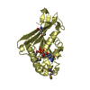
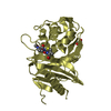
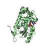
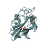
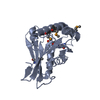
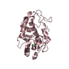
| Entry | Database: PDB / ID: 3ffv | ||||||
|---|---|---|---|---|---|---|---|
| Title | Crystal Structure Analysis of Syd | ||||||
 Components Components | Protein syd | ||||||
 Keywords Keywords |  PROTEIN BINDING / Membrane / Translocon / SecYEG / Syd / Nanodisc / Cell inner membrane / Cell membrane PROTEIN BINDING / Membrane / Translocon / SecYEG / Syd / Nanodisc / Cell inner membrane / Cell membrane | ||||||
| Function / homology |  Function and homology information Function and homology informationregulation of protein-containing complex assembly / extrinsic component of cytoplasmic side of plasma membrane / cytoplasmSimilarity search - Function | ||||||
| Biological species |  Escherichia coli (E. coli) Escherichia coli (E. coli) | ||||||
| Method | X-RAY DIFFRACTION / SYNCHROTRON / SAD / Resolution: 2 Å | ||||||
 Authors Authors | Maurus, R. / Brayer, G.D. | ||||||
 Citation Citation | Journal: J.Biol.Chem. / Year: 2009 Title: Structure, Binding, and Activity of Syd, a SecY-interacting Protein Authors: Dalal, K. / Nguyen, N. / Alami, M. / Tan, J. / Moraes, T.F. / Lee, W.C. / Maurus, R. / Sligar, S.S. / Brayer, G.D. / Duong, F. | ||||||
| History |
|
- Structure visualization
| Structure viewer | Molecule: MolmilJmol/JSmol |
|---|
- Downloads & links
-Download
| PDBx/mmCIF format | 3ffv.cif.gz | 90.9 KB | Display | PDBx/mmCIF format |
|---|---|---|---|---|
| PDB format | pdb3ffv.ent.gz | 73.4 KB | Display | PDB format |
| PDBx/mmJSON format | 3ffv.json.gz | Tree view | PDBx/mmJSON format | |
| Others |  Other downloads Other downloads |
-Validation report
| Arichive directory | https://data.pdbj.org/pub/pdb/validation_reports/ff/3ffvftp://data.pdbj.org/pub/pdb/validation_reports/ff/3ffv | HTTPS FTP |
|---|
-Related structure data
| Similar structure data |
|---|


-Links
 PDBj
PDBj
- Assembly
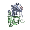
| Deposited unit | 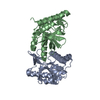
| ||||||||
|---|---|---|---|---|---|---|---|---|---|
| 1 | 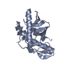
| ||||||||
| 2 | 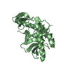
| ||||||||
| Unit cell |
|
-Components
| #1: Protein | Mass: 20729.420 Da / Num. of mol.: 2 / Source method: isolated from a natural source / Source: (natural) Escherichia coli (E. coli) / Strain: K12 / References: UniProt: P0A8U0#2: Water | ChemComp-HOH / | Water Mass: 18.015 Da / Num. of mol.: 510 / Source method: isolated from a natural source / Formula: H2O Mass: 18.015 Da / Num. of mol.: 510 / Source method: isolated from a natural source / Formula: H2O |
|---|
-Experimental details
-Experiment
| Experiment | Method: X-RAY DIFFRACTION / Number of used crystals: 2 |
|---|
- Sample preparation
| Crystal | Density Matthews: 2.71 Å3/Da / Density % sol: 54.65 % |
|---|---|
| Crystal grow | Temperature: 298 K / pH: 7 Details: 0.8-1.0 M sodium citrate, 0.2 M sodium chloride, 0.1 M Tris, pH 7.0, VAPOR DIFFUSION, HANGING DROP, temperature 298K |
-Data collection
| Diffraction |
| ||||||||||||||||||
|---|---|---|---|---|---|---|---|---|---|---|---|---|---|---|---|---|---|---|---|
| Diffraction source | Source: SYNCHROTRON / Site: SSRL  / Beamline: BL11-1 / Wavelength: 0.9788 / Beamline: BL11-1 / Wavelength: 0.9788 | ||||||||||||||||||
| Detector | Type: MARMOSAIC 325 MM CCD MARMOSAIC 325 MM CCD / Detector: CCD / Date: Jun 9, 2007 Details: FLAT MIRROR (VERTICAL FOCUSING); SINGLE CRYSTAL SI(111) BENT MONOCHROMATOR (HO RIZONTAL FOCUSING); FLAT MIRROR (VERTICAL FOCUSING); SINGLE CRYSTAL SI(111) BENT MONOCHROMATOR (HORIZONTAL FOCUSING) | ||||||||||||||||||
| Radiation |
| ||||||||||||||||||
| Radiation wavelength | Wavelength: 0.9788 Å / Relative weight: 1 | ||||||||||||||||||
| Reflection | Resolution: 2→50 Å / Num. obs: 29401 / % possible obs: 99.8 % / Observed criterion σ(I): 0 / Redundancy: 5.6 % / Rmerge(I) obs: 0.068 | ||||||||||||||||||
| Reflection shell | Resolution: 2→2.11 Å / Redundancy: 5.4 % / Rmerge(I) obs: 0.151 / Mean I/σ(I) obs: 8.8 / % possible all: 99.9 |
- Processing
| Software |
| |||||||||||||||||||||||||
|---|---|---|---|---|---|---|---|---|---|---|---|---|---|---|---|---|---|---|---|---|---|---|---|---|---|---|
| Refinement | Method to determine structure: SAD / Resolution: 2→41.9 Å / Isotropic thermal model: ISOTROPIC / σ(F): 0 / Stereochemistry target values: ENGH & HUBER
| |||||||||||||||||||||||||
| Displacement parameters | Biso mean: 25 Å2
| |||||||||||||||||||||||||
| Refinement step | Cycle: LAST / Resolution: 2→41.9 Å
|
