 Movie
Movie Controller


+ Open data

- Basic information
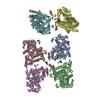
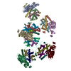
| Entry | Database: PDB / ID: 5mjs | ||||||
|---|---|---|---|---|---|---|---|
| Title | S. pombe microtubule copolymerized with GTP and Mal3-143 | ||||||
 Components Components |
| ||||||
 Keywords Keywords |  STRUCTURAL PROTEIN / Schizosaccharomyces pombe microtubules STRUCTURAL PROTEIN / Schizosaccharomyces pombe microtubules | ||||||
| Function / homology |  Function and homology information Function and homology informationmitotic spindle pole body duplication / Sealing of the nuclear envelope (NE) by ESCRT-III / Cilium Assembly / dynein-driven meiotic oscillatory nuclear movement / post-anaphase array microtubule end / Platelet degranulation / RHO GTPases activate IQGAPs / COPI-mediated anterograde transport / nuclear migration involved in conjugation with cellular fusion / cell cortex of cell tip ...mitotic spindle pole body duplication / Sealing of the nuclear envelope (NE) by ESCRT-III / Cilium Assembly / dynein-driven meiotic oscillatory nuclear movement / post-anaphase array microtubule end / Platelet degranulation / RHO GTPases activate IQGAPs / COPI-mediated anterograde transport / nuclear migration involved in conjugation with cellular fusion / cell cortex of cell tip / cortical microtubule / nuclear migration by microtubule mediated pushing forces / Neutrophil degranulation / mitotic spindle astral microtubule / karyogamy involved in conjugation with cellular fusion / nuclear division / mitotic spindle pole body / mitotic spindle elongation / nuclear microtubule / mitotic spindle midzone / astral microtubule / protein localization to microtubule / microtubule plus-end / cytoskeletal anchor activity / attachment of mitotic spindle microtubules to kinetochore / microtubule plus-end binding / microtubule organizing center / microtubule lateral binding / intracellular distribution of mitochondria / ATPase activator activity / spindle assembly / regulation of microtubule polymerization or depolymerization / spindle midzone / cytoplasmic microtubule / cytoplasmic microtubule organization / molecular condensate scaffold activity / spindle microtubule / Hydrolases; Acting on acid anhydrides; Acting on GTP to facilitate cellular and subcellular movement / structural constituent of cytoskeleton / spindle / microtubule cytoskeleton organization / microtubule cytoskeleton / mitotic cell cycle / microtubule binding / microtubule / hydrolase activity / cell division / response to antibiotic / GTPase activity / GTP binding / metal ion binding / nucleus / cytosol / cytoplasmSimilarity search - Function | ||||||
| Biological species |  Schizosaccharomyces pombe (fission yeast) Schizosaccharomyces pombe (fission yeast) | ||||||
| Method | ELECTRON MICROSCOPY / single particle reconstruction / cryo EM / Resolution: 4.6 Å | ||||||
 Authors Authors | von Loeffelholz, O. / Moores, C. | ||||||
| Funding support |  United Kingdom, 1items United Kingdom, 1items
| ||||||
 Citation Citation | Journal: Nat Commun / Year: 2017 Title: Nucleotide- and Mal3-dependent changes in fission yeast microtubules suggest a structural plasticity view of dynamics. Authors: Ottilie von Loeffelholz / Neil A Venables / Douglas Robert Drummond / Miho Katsuki / Robert Cross / Carolyn A Moores /   Abstract: Using cryo-electron microscopy, we characterize the architecture of microtubules assembled from Schizosaccharomyces pombe tubulin, in the presence and absence of their regulatory partner Mal3. Cryo- ...Using cryo-electron microscopy, we characterize the architecture of microtubules assembled from Schizosaccharomyces pombe tubulin, in the presence and absence of their regulatory partner Mal3. Cryo-electron tomography reveals that microtubules assembled from S. pombe tubulin have predominantly B-lattice interprotofilament contacts, with protofilaments skewed around the microtubule axis. Copolymerization with Mal3 favors 13 protofilament microtubules with reduced protofilament skew, indicating that Mal3 adjusts interprotofilament interfaces. A 4.6-Å resolution structure of microtubule-bound Mal3 shows that Mal3 makes a distinctive footprint on the S. pombe microtubule lattice and that unlike mammalian microtubules, S. pombe microtubules do not show the longitudinal lattice compaction associated with EB protein binding and GTP hydrolysis. Our results firmly support a structural plasticity view of microtubule dynamics in which microtubule lattice conformation is sensitive to a variety of effectors and differently so for different tubulins. | ||||||
| History |
|
- Structure visualization
| Movie |
Movie viewer |
|---|---|
| Structure viewer | Molecule: MolmilJmol/JSmol |
- Downloads & links
-Download
| PDBx/mmCIF format | 5mjs.cif.gz | 604 KB | Display | PDBx/mmCIF format |
|---|---|---|---|---|
| PDB format | pdb5mjs.ent.gz | 516.7 KB | Display | PDB format |
| PDBx/mmJSON format | 5mjs.json.gz | Tree view | PDBx/mmJSON format | |
| Others |  Other downloads Other downloads |
-Validation report
| Arichive directory | https://data.pdbj.org/pub/pdb/validation_reports/mj/5mjsftp://data.pdbj.org/pub/pdb/validation_reports/mj/5mjs | HTTPS FTP |
|---|
-Related structure data
| Related structure data |  3522MC M: map data used to model this data C: citing same article ( |
|---|---|
| Similar structure data |


-Links
 PDBj
PDBj


- Assembly
| Deposited unit | 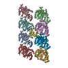
|
|---|---|
| 1 |
|
-Components
| #1: Protein | Mass: 47191.031 Da / Num. of mol.: 4 Source method: isolated from a genetically manipulated source Source: (gene. exp.) Schizosaccharomyces pombe (strain 972 / ATCC 24843) (yeast)Strain: 972 / ATCC 24843 / Gene: nda3, alp12, SPBC26H8.07c / Production host: Schizosaccharomyces pombe (fission yeast) / References: UniProt: P05219#2: Protein | | Mass: 16668.900 Da / Num. of mol.: 1 Source method: isolated from a genetically manipulated source Source: (gene. exp.) Schizosaccharomyces pombe (strain 972 / ATCC 24843) (yeast)Strain: 972 / ATCC 24843 / Gene: mal3, SPAC18G6.15 / Production host:  Escherichia coli (E. coli) / References: UniProt: Q10113 Escherichia coli (E. coli) / References: UniProt: Q10113#3: Protein | Mass: 49813.832 Da / Num. of mol.: 4 Source method: isolated from a genetically manipulated source Source: (gene. exp.) Schizosaccharomyces pombe (strain 972 / ATCC 24843) (yeast)Strain: 972 / ATCC 24843 / Gene: nda2, SPBC16A3.15c / Production host: Schizosaccharomyces pombe (fission yeast) / References: UniProt: P04688#4: Chemical | ChemComp-GDP / Guanosine diphosphate  Type: RNA linking / Mass: 443.201 Da / Num. of mol.: 4 Type: RNA linking / Mass: 443.201 Da / Num. of mol.: 4Source method: isolated from a genetically manipulated source Formula: C10H15N5O11P2 Source: (gene. exp.) Schizosaccharomyces pombe (fission yeast)Production host: Schizosaccharomyces pombe (fission yeast) / Comment: GDP, energy-carrying molecule*YM#5: Chemical | ChemComp-GTP / Guanosine triphosphate  Mass: 523.180 Da / Num. of mol.: 4 / Source method: obtained synthetically / Formula: C10H16N5O14P3 Mass: 523.180 Da / Num. of mol.: 4 / Source method: obtained synthetically / Formula: C10H16N5O14P3Source: (gene. exp.) Schizosaccharomyces pombe (fission yeast)Production host: Schizosaccharomyces pombe (fission yeast) / Comment: GTP, energy-carrying molecule*YM |
|---|
-Experimental details
-Experiment
| Experiment | Method: ELECTRON MICROSCOPY |
|---|---|
| EM experiment | Aggregation state: HELICAL ARRAY / 3D reconstruction method: single particle reconstruction |
- Sample preparation
| Component | Name: Microtubule decorated with Mal3 / Type: COMPLEX / Entity ID: #1-#3 / Source: RECOMBINANT |
|---|---|
| Molecular weight | Experimental value: NO |
| Source (natural) | Organism: Schizosaccharomyces pombe (fission yeast) |
| Source (recombinant) | Organism: Schizosaccharomyces pombe (fission yeast) |
| Buffer solution | pH: 6.5 |
| Specimen | Conc.: 2 mg/ml / Embedding applied: NO / Shadowing applied: NO / Staining applied: NO / Vitrification applied: YES |
| Specimen support | Grid material: COPPER / Grid mesh size: 400 divisions/in. / Grid type: Quantifoil R2/2 |
| Vitrification | Instrument: FEI VITROBOT MARK IV / Cryogen name: ETHANE / Humidity: 100 % |
- Electron microscopy imaging
| Experimental equipment |  Model: Tecnai Polara / Image courtesy: FEI Company |
|---|---|
| Microscopy | Model: FEI POLARA 300 |
| Electron gun | Electron source: FIELD EMISSION GUN / Accelerating voltage: 300 kV / Illumination mode: OTHER |
| Electron lens | Mode: BRIGHT FIELDBright-field microscopy |
| Image recording | Electron dose: 30 e/Å2 / Detector mode: COUNTING / Film or detector model: GATAN K2 SUMMIT (4k x 4k) |
- Processing
| Software | Name: PHENIX / Version: 1.11.1_2575: / Classification: refinement | ||||||||||||||||||||||||||||||||
|---|---|---|---|---|---|---|---|---|---|---|---|---|---|---|---|---|---|---|---|---|---|---|---|---|---|---|---|---|---|---|---|---|---|
| EM software |
| ||||||||||||||||||||||||||||||||
| CTF correction | Type: PHASE FLIPPING AND AMPLITUDE CORRECTION | ||||||||||||||||||||||||||||||||
| 3D reconstruction | Resolution: 4.6 Å / Resolution method: FSC 0.143 CUT-OFF / Num. of particles: 12763 / Symmetry type: POINT | ||||||||||||||||||||||||||||||||
| Atomic model building | Protocol: BACKBONE TRACE / Space: REAL | ||||||||||||||||||||||||||||||||
| Refine LS restraints |
|
