 Movie
Movie Controller

[English] 日本語
 Yorodumi
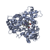
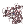
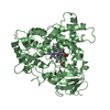
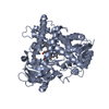
- PDB-3qu8: Crystal structure of a human cytochrome P450 2B6 (Y226H/K262R) in... -
+ Open data

- Basic information
| Entry | Database: PDB / ID: 3qu8 | ||||||
|---|---|---|---|---|---|---|---|
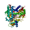
| Title | Crystal structure of a human cytochrome P450 2B6 (Y226H/K262R) in complex with the inhibitor 4-(4-Nitrobenzyl)pyridine. | ||||||
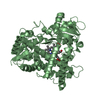
 Components Components | Cytochrome P450 2B6 | ||||||
 Keywords Keywords | OXIDOREDUCTASE/OXIDOREDUCTASE INHIBITOR /  P450 / CYTOCHROME P450 2B6 / MONOOXYGENASE / MEMBRANE PROTEIN / CYP2B6 / ENDOPLASMIC RETICULUM / HEME / IRON / MEMBRANE / METAL BINDING / MICROSOME / PHOSPHOPROTEIN / OXIDOREDUCTASE-OXIDOREDUCTASE INHIBITOR complex P450 / CYTOCHROME P450 2B6 / MONOOXYGENASE / MEMBRANE PROTEIN / CYP2B6 / ENDOPLASMIC RETICULUM / HEME / IRON / MEMBRANE / METAL BINDING / MICROSOME / PHOSPHOPROTEIN / OXIDOREDUCTASE-OXIDOREDUCTASE INHIBITOR complex | ||||||
| Function / homology |  Function and homology information Function and homology informationtestosterone 16-alpha-hydroxylase activity / testosterone 16-beta-hydroxylase activity / Fatty acids / Oxidoreductases; Acting on paired donors, with incorporation or reduction of molecular oxygen; With NADH or NADPH as one donor, and incorporation of one atom of oxygen into the other donor / cellular ketone metabolic process / arachidonic acid epoxygenase activity / CYP2E1 reactions / epoxygenase P450 pathway / estrogen 2-hydroxylase activity / anandamide 8,9 epoxidase activity ...testosterone 16-alpha-hydroxylase activity / testosterone 16-beta-hydroxylase activity / Fatty acids / Oxidoreductases; Acting on paired donors, with incorporation or reduction of molecular oxygen; With NADH or NADPH as one donor, and incorporation of one atom of oxygen into the other donor / cellular ketone metabolic process / arachidonic acid epoxygenase activity / CYP2E1 reactions / epoxygenase P450 pathway / estrogen 2-hydroxylase activity / anandamide 8,9 epoxidase activity / anandamide 11,12 epoxidase activity / anandamide 14,15 epoxidase activity / Xenobiotics / oxidoreductase activity, acting on paired donors, with incorporation or reduction of molecular oxygen, reduced flavin or flavoprotein as one donor, and incorporation of one atom of oxygen / Phase I - Functionalization of compounds / steroid metabolic process / xenobiotic catabolic process / xenobiotic metabolic process / monooxygenase activity / iron ion binding / intracellular membrane-bounded organelle / heme binding / endoplasmic reticulum membrane / cytoplasmSimilarity search - Function | ||||||
| Biological species |  Homo sapiens (human) Homo sapiens (human) | ||||||
| Method | X-RAY DIFFRACTION / SYNCHROTRON / MOLECULAR REPLACEMENT / Resolution: 2.8 Å | ||||||
 Authors Authors | Shah, M.B. / Pascual, J. / Stout, C.D. / Halpert, J.R. | ||||||
 Citation Citation | Journal: Mol.Pharmacol. / Year: 2011 Title: Structures of Cytochrome P450 2B6 Bound to 4-Benzylpyridine and 4-(4-Nitrobenzyl)pyridine: Insight into Inhibitor Binding and Rearrangement of Active Site Side Chains. Authors: Shah, M.B. / Pascual, J. / Zhang, Q. / Stout, C.D. / Halpert, J.R. | ||||||
| History |
| ||||||
| Remark 999 | THE RESIDUES 3-21 (LSVLLFLALLTGLLLLLVQ) OF CORRESPONDING DATABASE REFERENCE SEQUENCE (UNP P20813) ...THE RESIDUES 3-21 (LSVLLFLALLTGLLLLLVQ) OF CORRESPONDING DATABASE REFERENCE SEQUENCE (UNP P20813) IS DELETED IN THIS STRUCTURE. |
- Structure visualization
| Structure viewer | Molecule: MolmilJmol/JSmol |
|---|
- Downloads & links
-Download
| PDBx/mmCIF format | 3qu8.cif.gz | 557.1 KB | Display | PDBx/mmCIF format |
|---|---|---|---|---|
| PDB format | pdb3qu8.ent.gz | 462.1 KB | Display | PDB format |
| PDBx/mmJSON format | 3qu8.json.gz | Tree view | PDBx/mmJSON format | |
| Others |  Other downloads Other downloads |
-Validation report
| Arichive directory | https://data.pdbj.org/pub/pdb/validation_reports/qu/3qu8ftp://data.pdbj.org/pub/pdb/validation_reports/qu/3qu8 | HTTPS FTP |
|---|
-Related structure data
| Related structure data |  3qoaC  3ibdS S: Starting model for refinement C: citing same article ( |
|---|---|
| Similar structure data |


-Links
 PDBj
PDBj

- Assembly
| Deposited unit | 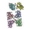
| |||||||||||||||||||||||||||||||||||||||||||||||||||||||||||||||
|---|---|---|---|---|---|---|---|---|---|---|---|---|---|---|---|---|---|---|---|---|---|---|---|---|---|---|---|---|---|---|---|---|---|---|---|---|---|---|---|---|---|---|---|---|---|---|---|---|---|---|---|---|---|---|---|---|---|---|---|---|---|---|---|---|
| 1 | 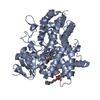
| |||||||||||||||||||||||||||||||||||||||||||||||||||||||||||||||
| 2 | 
| |||||||||||||||||||||||||||||||||||||||||||||||||||||||||||||||
| 3 | 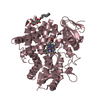
| |||||||||||||||||||||||||||||||||||||||||||||||||||||||||||||||
| 4 | 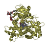
| |||||||||||||||||||||||||||||||||||||||||||||||||||||||||||||||
| 5 | 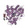
| |||||||||||||||||||||||||||||||||||||||||||||||||||||||||||||||
| 6 | 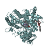
| |||||||||||||||||||||||||||||||||||||||||||||||||||||||||||||||
| Unit cell |
| |||||||||||||||||||||||||||||||||||||||||||||||||||||||||||||||
| Noncrystallographic symmetry (NCS) | NCS domain:
NCS domain segments:
|
-Components
| #1: Protein | Mass: 54646.004 Da / Num. of mol.: 6 / Fragment: Cytochrome P450 2B6, residues 3-21 deleted Mutation: E2A, R22K, H23K, P24T, N25S, T26S, H27K, D28G, R29K, Y226H, K262R Source method: isolated from a genetically manipulated source Source: (gene. exp.) Homo sapiens (human) / Gene: CYP2B6 / Plasmid: pKK / Production host:  Escherichia coli (E. coli) / Strain (production host): JM109 / References: UniProt: P20813, unspecific monooxygenase Escherichia coli (E. coli) / Strain (production host): JM109 / References: UniProt: P20813, unspecific monooxygenase#2: Chemical | ChemComp-HEM / Heme B  Mass: 616.487 Da / Num. of mol.: 6 / Source method: obtained synthetically / Formula: C34H32FeN4O4 Mass: 616.487 Da / Num. of mol.: 6 / Source method: obtained synthetically / Formula: C34H32FeN4O4#3: Chemical | ChemComp-3QU /   Mass: 214.220 Da / Num. of mol.: 5 / Source method: obtained synthetically / Formula: C12H10N2O2 Mass: 214.220 Da / Num. of mol.: 5 / Source method: obtained synthetically / Formula: C12H10N2O2#4: Chemical | ChemComp-CM5 / 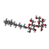  Mass: 494.573 Da / Num. of mol.: 8 / Source method: obtained synthetically / Formula: C23H42O11 / Comment: detergent*YM Mass: 494.573 Da / Num. of mol.: 8 / Source method: obtained synthetically / Formula: C23H42O11 / Comment: detergent*YM#5: Water | ChemComp-HOH / | Water Mass: 18.015 Da / Num. of mol.: 150 / Source method: isolated from a natural source / Formula: H2O Mass: 18.015 Da / Num. of mol.: 150 / Source method: isolated from a natural source / Formula: H2O |
|---|
-Experimental details
-Experiment
| Experiment | Method: X-RAY DIFFRACTION / Number of used crystals: 1 |
|---|
- Sample preparation
| Crystal | Density Matthews: 2.74 Å3/Da / Density % sol: 55.06 % |
|---|---|
| Crystal grow | Temperature: 291 K / pH: 7 Details: 0.15 M DL-Malic Acid, 20 % w/v PEG 3350, pH 7, VAPOR DIFFUSION, SITTING DROP, temperature 291K |
-Data collection
| Diffraction | Mean temperature: 100 K |
|---|---|
| Diffraction source | Source: SYNCHROTRON / Site: SSRL  / Beamline: BL11-1 / Wavelength: 0.98 / Beamline: BL11-1 / Wavelength: 0.98 |
| Detector | Type: MARMOSAIC 325 mm CCD / Detector: CCD / Date: Dec 11, 2010 |
| Radiation | Monochromator: SI(111) / Protocol: SINGLE WAVELENGTH / Monochromatic (M) / Laue (L): M / Scattering type: x-ray |
| Radiation wavelength | Wavelength: 0.98 Å / Relative weight: 1 |
| Reflection | Resolution: 2.8→84.6 Å / Num. obs: 76085 / % possible obs: 88.9 % / Observed criterion σ(I): 0 / Redundancy: 2.6 % / Biso Wilson estimate: 52.67 Å2 / Rmerge(I) obs: 0.063 / Rsym value: 0.063 / Net I/σ(I): 9.6 |
| Reflection shell | Resolution: 2.8→2.95 Å / Redundancy: 2.4 % / Rmerge(I) obs: 0.368 / Mean I/σ(I) obs: 2.1 / Rsym value: 0.368 / % possible all: 78.6 |
- Processing
| Software |
| ||||||||||||||||||||||||||||||||||||||||||||||||||||||||||||||||||||||||||||||||||||||||||||||||||||||||||||||||||||||||||||||||||||||||||||||||||||||||||||||||||||||||||
|---|---|---|---|---|---|---|---|---|---|---|---|---|---|---|---|---|---|---|---|---|---|---|---|---|---|---|---|---|---|---|---|---|---|---|---|---|---|---|---|---|---|---|---|---|---|---|---|---|---|---|---|---|---|---|---|---|---|---|---|---|---|---|---|---|---|---|---|---|---|---|---|---|---|---|---|---|---|---|---|---|---|---|---|---|---|---|---|---|---|---|---|---|---|---|---|---|---|---|---|---|---|---|---|---|---|---|---|---|---|---|---|---|---|---|---|---|---|---|---|---|---|---|---|---|---|---|---|---|---|---|---|---|---|---|---|---|---|---|---|---|---|---|---|---|---|---|---|---|---|---|---|---|---|---|---|---|---|---|---|---|---|---|---|---|---|---|---|---|---|---|---|
| Refinement | Method to determine structure: MOLECULAR REPLACEMENT Starting model: 3IBD Resolution: 2.8→50.51 Å / Cor.coef. Fo:Fc: 0.917 / Cor.coef. Fo:Fc free: 0.884 / SU B: 16.814 / SU ML: 0.33 / Cross valid method: THROUGHOUT / σ(F): 0 / ESU R Free: 0.433 / Stereochemistry target values: MAXIMUM LIKELIHOOD / Details: HYDROGENS HAVE BEEN ADDED IN THE RIDING POSITIONS
| ||||||||||||||||||||||||||||||||||||||||||||||||||||||||||||||||||||||||||||||||||||||||||||||||||||||||||||||||||||||||||||||||||||||||||||||||||||||||||||||||||||||||||
| Solvent computation | Ion probe radii: 0.8 Å / Shrinkage radii: 0.8 Å / VDW probe radii: 1.4 Å / Solvent model: MASK | ||||||||||||||||||||||||||||||||||||||||||||||||||||||||||||||||||||||||||||||||||||||||||||||||||||||||||||||||||||||||||||||||||||||||||||||||||||||||||||||||||||||||||
| Displacement parameters | Biso mean: 52.67 Å2
| ||||||||||||||||||||||||||||||||||||||||||||||||||||||||||||||||||||||||||||||||||||||||||||||||||||||||||||||||||||||||||||||||||||||||||||||||||||||||||||||||||||||||||
| Refinement step | Cycle: LAST / Resolution: 2.8→50.51 Å
| ||||||||||||||||||||||||||||||||||||||||||||||||||||||||||||||||||||||||||||||||||||||||||||||||||||||||||||||||||||||||||||||||||||||||||||||||||||||||||||||||||||||||||
| Refine LS restraints |
| ||||||||||||||||||||||||||||||||||||||||||||||||||||||||||||||||||||||||||||||||||||||||||||||||||||||||||||||||||||||||||||||||||||||||||||||||||||||||||||||||||||||||||
| Refine LS restraints NCS | Ens-ID: 1 / Refine-ID: X-RAY DIFFRACTION
| ||||||||||||||||||||||||||||||||||||||||||||||||||||||||||||||||||||||||||||||||||||||||||||||||||||||||||||||||||||||||||||||||||||||||||||||||||||||||||||||||||||||||||
| LS refinement shell | Resolution: 2.8→2.88 Å / Total num. of bins used: 20
|
