 Movie
Movie Controller


+ Open data

- Basic information
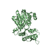
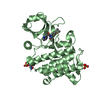
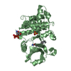
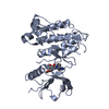
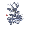
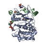
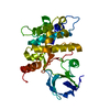
| Entry | Database: PDB / ID: 5mgr | |||||||||
|---|---|---|---|---|---|---|---|---|---|---|
| Title | Human receptor NKR-P1 in glycosylated form, extracellular domain | |||||||||
 Components Components | Killer cell lectin-like receptor subfamily B member 1 | |||||||||
 Keywords Keywords |  IMMUNE SYSTEM / receptor / CTL fold / natural killer cell IMMUNE SYSTEM / receptor / CTL fold / natural killer cell | |||||||||
| Function / homology |  Function and homology information Function and homology informationregulation of natural killer cell mediated cytotoxicity / Immunoregulatory interactions between a Lymphoid and a non-Lymphoid cell / transmembrane signaling receptor activity / signaling receptor activity / carbohydrate binding / cell surface receptor signaling pathway / cell surface / plasma membraneSimilarity search - Function | |||||||||
| Biological species |  Homo sapiens (human) Homo sapiens (human) | |||||||||
| Method | X-RAY DIFFRACTION / SYNCHROTRON / MOLECULAR REPLACEMENT / Resolution: 1.8 Å | |||||||||
 Authors Authors | Skalova, T. / Blaha, J. / Stransky, J. / Koval, T. / Hasek, J. / Yuguang, Z. / Harlos, K. / Vanek, O. / Dohnalek, J. | |||||||||
| Funding support |  Czech Republic, 2items Czech Republic, 2items
| |||||||||
 Citation Citation | Journal: Nat Commun / Year: 2022 Title: Structure of the human NK cell NKR-P1:LLT1 receptor:ligand complex reveals clustering in the immune synapse. Authors: Blaha, J. / Skalova, T. / Kalouskova, B. / Skorepa, O. / Cmunt, D. / Grobarova, V. / Pazicky, S. / Polachova, E. / Abreu, C. / Stransky, J. / Koval, T. / Duskova, J. / Zhao, Y. / Harlos, K. ...Authors: Blaha, J. / Skalova, T. / Kalouskova, B. / Skorepa, O. / Cmunt, D. / Grobarova, V. / Pazicky, S. / Polachova, E. / Abreu, C. / Stransky, J. / Koval, T. / Duskova, J. / Zhao, Y. / Harlos, K. / Hasek, J. / Dohnalek, J. / Vanek, O. | |||||||||
| History |
|
- Structure visualization
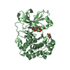
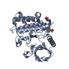
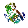
| Structure viewer | Molecule: MolmilJmol/JSmol |
|---|
- Downloads & links
-Download
| PDBx/mmCIF format | 5mgr.cif.gz | 77.6 KB | Display | PDBx/mmCIF format |
|---|---|---|---|---|
| PDB format | pdb5mgr.ent.gz | 56.5 KB | Display | PDB format |
| PDBx/mmJSON format | 5mgr.json.gz | Tree view | PDBx/mmJSON format | |
| Others |  Other downloads Other downloads |
-Validation report
| Arichive directory | https://data.pdbj.org/pub/pdb/validation_reports/mg/5mgrftp://data.pdbj.org/pub/pdb/validation_reports/mg/5mgr | HTTPS FTP |
|---|
-Related structure data
| Related structure data | 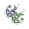 5mgsC 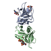 5mgtC 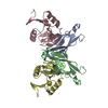 3ff7S S: Starting model for refinement C: citing same article ( |
|---|---|
| Similar structure data |
-Links
 PDBj
PDBj


- Assembly
| Deposited unit | 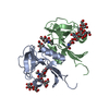
| ||||||||
|---|---|---|---|---|---|---|---|---|---|
| 1 |
| ||||||||
| Unit cell |
|
-Components
| #1: Protein | Mass: 17108.312 Da / Num. of mol.: 2 Source method: isolated from a genetically manipulated source Source: (gene. exp.) Homo sapiens (human) / Cell: lymphocytes, natural killer cell / Gene: KLRB1, CLEC5B, NKRP1A / Plasmid: pOPINGGTneo / Cell line (production host): HEK293S GnTI- / Organ (production host): Human embryonic kidney / Production host: Homo sapiens (human) / References: UniProt: Q12918#2: Polysaccharide | alpha-D-mannopyranose-(1-3)-[alpha-D-mannopyranose-(1-6)]alpha-D-mannopyranose-(1-6)-[alpha-D- ...alpha-D-mannopyranose-(1-3)-[alpha-D-mannopyranose-(1-6)]alpha-D-mannopyranose-(1-6)-[alpha-D-mannopyranose-(1-3)]beta-D-mannopyranose-(1-4)-2-acetamido-2-deoxy-beta-D-glucopyranose-(1-4)-2-acetamido-2-deoxy-beta-D-glucopyranose | / Mass: 1235.105 Da / Num. of mol.: 1Source method: isolated from a genetically manipulated source #3: Polysaccharide | alpha-D-mannopyranose-(1-3)-[alpha-D-mannopyranose-(1-6)]beta-D-mannopyranose-(1-4)-2-acetamido-2- ...alpha-D-mannopyranose-(1-3)-[alpha-D-mannopyranose-(1-6)]beta-D-mannopyranose-(1-4)-2-acetamido-2-deoxy-beta-D-glucopyranose-(1-4)-2-acetamido-2-deoxy-beta-D-glucopyranose | / Mass: 910.823 Da / Num. of mol.: 1Source method: isolated from a genetically manipulated source #4: Sugar | N-Acetylglucosamine  Type: D-saccharide, beta linking / Mass: 221.208 Da / Num. of mol.: 2 Type: D-saccharide, beta linking / Mass: 221.208 Da / Num. of mol.: 2Source method: isolated from a genetically manipulated source Formula: C8H15NO6 #5: Water | ChemComp-HOH / | Water Mass: 18.015 Da / Num. of mol.: 225 / Source method: isolated from a natural source / Formula: H2O Mass: 18.015 Da / Num. of mol.: 225 / Source method: isolated from a natural source / Formula: H2O |
|---|
-Experimental details
-Experiment
| Experiment | Method: X-RAY DIFFRACTION / Number of used crystals: 1 |
|---|
- Sample preparation
| Crystal | Density Matthews: 2.32 Å3/Da / Density % sol: 47 % Description: A hexagonal crystal with dimensions 150, 150, 20 um |
|---|---|
| Crystal grow | Temperature: 294 K / Method: vapor diffusion, sitting drop / pH: 7.2 Details: 20% (w/v) PEG 3350, 200 mM di-sodium tartrate pH 7.2 PROTEIN CONCENTRATION 20 MG/ML |
-Data collection
| Diffraction | Mean temperature: 100 K |
|---|---|
| Diffraction source | Source: SYNCHROTRON / Site: Diamond  / Beamline: I03 / Wavelength: 0.97625 Å / Beamline: I03 / Wavelength: 0.97625 Å |
| Detector | Type: DECTRIS PILATUS3 6M / Detector: PIXEL / Date: May 7, 2015 |
| Radiation | Protocol: SINGLE WAVELENGTH / Monochromatic (M) / Laue (L): M / Scattering type: x-ray |
| Radiation wavelength | Wavelength: 0.97625 Å / Relative weight: 1 |
| Reflection | Resolution: 1.8→43.3 Å / Num. obs: 32555 / % possible obs: 100 % / Observed criterion σ(F): 0 / Observed criterion σ(I): -3.7 / Redundancy: 39.5 % / Biso Wilson estimate: 25.3 Å2 / CC1/2: 1 / Rmerge(I) obs: 0.061 / Net I/σ(I): 41 |
| Reflection shell | Resolution: 1.8→1.84 Å / Redundancy: 40.5 % / Rmerge(I) obs: 0.637 / Mean I/σ(I) obs: 7.4 / Num. unique all: 1900 / CC1/2: 0.975 / % possible all: 100 |
- Processing
| Software |
| ||||||||||||||||||||||||||||||||||||||||||||||||||||||||||||||||||||||||||||||||||||||||||||||||||||||||||||||||||||||||||||||||||||||||||||||||||||||||||||||||||||||||||||||||||||||
|---|---|---|---|---|---|---|---|---|---|---|---|---|---|---|---|---|---|---|---|---|---|---|---|---|---|---|---|---|---|---|---|---|---|---|---|---|---|---|---|---|---|---|---|---|---|---|---|---|---|---|---|---|---|---|---|---|---|---|---|---|---|---|---|---|---|---|---|---|---|---|---|---|---|---|---|---|---|---|---|---|---|---|---|---|---|---|---|---|---|---|---|---|---|---|---|---|---|---|---|---|---|---|---|---|---|---|---|---|---|---|---|---|---|---|---|---|---|---|---|---|---|---|---|---|---|---|---|---|---|---|---|---|---|---|---|---|---|---|---|---|---|---|---|---|---|---|---|---|---|---|---|---|---|---|---|---|---|---|---|---|---|---|---|---|---|---|---|---|---|---|---|---|---|---|---|---|---|---|---|---|---|---|---|
| Refinement | Method to determine structure: MOLECULAR REPLACEMENT Starting model: 3ff7 Resolution: 1.8→43.3 Å / Cor.coef. Fo:Fc: 0.966 / SU B: 1.787 / SU ML: 0.055 / Cross valid method: FREE R-VALUE / ESU R: 0.1 / Details: HYDROGENS HAVE BEEN ADDED IN THE RIDING POSITIONS
| ||||||||||||||||||||||||||||||||||||||||||||||||||||||||||||||||||||||||||||||||||||||||||||||||||||||||||||||||||||||||||||||||||||||||||||||||||||||||||||||||||||||||||||||||||||||
| Solvent computation | Ion probe radii: 0.8 Å / Shrinkage radii: 0.8 Å / VDW probe radii: 1.2 Å | ||||||||||||||||||||||||||||||||||||||||||||||||||||||||||||||||||||||||||||||||||||||||||||||||||||||||||||||||||||||||||||||||||||||||||||||||||||||||||||||||||||||||||||||||||||||
| Displacement parameters | Biso mean: 33.5 Å2
| ||||||||||||||||||||||||||||||||||||||||||||||||||||||||||||||||||||||||||||||||||||||||||||||||||||||||||||||||||||||||||||||||||||||||||||||||||||||||||||||||||||||||||||||||||||||
| Refinement step | Cycle: 1 / Resolution: 1.8→43.3 Å
| ||||||||||||||||||||||||||||||||||||||||||||||||||||||||||||||||||||||||||||||||||||||||||||||||||||||||||||||||||||||||||||||||||||||||||||||||||||||||||||||||||||||||||||||||||||||
| Refine LS restraints |
|