 Movie
Movie Controller

[English] 日本語
 Yorodumi
- PDB-5mbz: Crystal Structure of Ser202Phe mutant of Human Prolidase with Mn ... -
+ Open data

- Basic information
| Entry | Database: PDB / ID: 5mbz | ||||||
|---|---|---|---|---|---|---|---|
| Title | Crystal Structure of Ser202Phe mutant of Human Prolidase with Mn ions and GlyPro ligand | ||||||
 Components Components | Xaa-Pro dipeptidase | ||||||
 Keywords Keywords |  HYDROLASE / prolidase / peptidase / hydrolysis / pita-bread / metalloenzyme / mutation HYDROLASE / prolidase / peptidase / hydrolysis / pita-bread / metalloenzyme / mutation | ||||||
| Function / homology |  Function and homology information Function and homology informationXaa-Pro dipeptidase / proline dipeptidase activity / negative regulation of programmed cell death / amino acid metabolic process / metalloaminopeptidase activity / collagen catabolic process / metallocarboxypeptidase activity / manganese ion binding / peptidase activity / proteolysis / extracellular exosomeSimilarity search - Function | ||||||
| Biological species |  Homo sapiens (human) Homo sapiens (human) | ||||||
| Method | X-RAY DIFFRACTION / SYNCHROTRON / MOLECULAR REPLACEMENT / molecular replacement / Resolution: 1.5 Å | ||||||
 Authors Authors | Wilk, P. / Mueller, U. / Dobbek, H. / Weiss, M.S. | ||||||
 Citation Citation | Journal: FEBS J. / Year: 2018 Title: Structural basis for prolidase deficiency disease mechanisms. Authors: Wilk, P. / Uehlein, M. / Piwowarczyk, R. / Dobbek, H. / Mueller, U. / Weiss, M.S. | ||||||
| History |
|
- Structure visualization
| Structure viewer | Molecule: MolmilJmol/JSmol |
|---|
- Downloads & links
-Download
| PDBx/mmCIF format | 5mbz.cif.gz | 444 KB | Display | PDBx/mmCIF format |
|---|---|---|---|---|
| PDB format | pdb5mbz.ent.gz | 364.1 KB | Display | PDB format |
| PDBx/mmJSON format | 5mbz.json.gz | Tree view | PDBx/mmJSON format | |
| Others |  Other downloads Other downloads |
-Validation report
| Arichive directory | https://data.pdbj.org/pub/pdb/validation_reports/mb/5mbzftp://data.pdbj.org/pub/pdb/validation_reports/mb/5mbz | HTTPS FTP |
|---|
-Related structure data
| Related structure data | 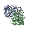 5mbyC 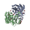 5mc0C 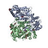 5mc1C 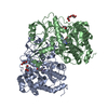 5mc2C  5mc3C 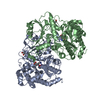 5mc4C 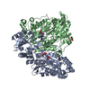 5mc5C 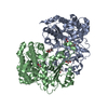 6h2pC 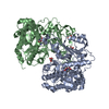 6h2qC 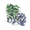 5m4jS S: Starting model for refinement C: citing same article ( |
|---|---|
| Similar structure data | |
| Experimental dataset #1 | Data reference: 10.18430/m35mbz / Data set type: diffraction image data |


-Links
 PDBj
PDBj


- Assembly
| Deposited unit | 
| ||||||||
|---|---|---|---|---|---|---|---|---|---|
| 1 |
| ||||||||
| Unit cell |
| ||||||||
| Components on special symmetry positions |
|
-Components
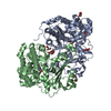
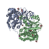
-Protein , 1 types, 2 molecules AB
| #1: Protein | Mass: 53859.191 Da / Num. of mol.: 2 / Mutation: S202F Source method: isolated from a genetically manipulated source Source: (gene. exp.) Homo sapiens (human) / Gene: PEPD, PRD / Plasmid: pET28a / Production host:  Escherichia coli BL21 (bacteria) / Variant (production host): Rosetta / References: UniProt: P12955, Xaa-Pro dipeptidase Escherichia coli BL21 (bacteria) / Variant (production host): Rosetta / References: UniProt: P12955, Xaa-Pro dipeptidase |
|---|
-Non-polymers , 8 types, 1143 molecules 


| #2: Chemical | ChemComp-MN /  Mass: 54.938 Da / Num. of mol.: 4 / Source method: obtained synthetically / Formula: Mn Mass: 54.938 Da / Num. of mol.: 4 / Source method: obtained synthetically / Formula: Mn#3: Chemical | Hydroxide Mass: 17.007 Da / Num. of mol.: 2 / Source method: obtained synthetically / Formula: HO Mass: 17.007 Da / Num. of mol.: 2 / Source method: obtained synthetically / Formula: HO#4: Chemical | Glycine Type: peptide linking / Mass: 75.067 Da / Num. of mol.: 2 / Source method: obtained synthetically / Formula: C2H5NO2 / Details: Substrate / Source: (synth.) Homo sapiens (human) Type: peptide linking / Mass: 75.067 Da / Num. of mol.: 2 / Source method: obtained synthetically / Formula: C2H5NO2 / Details: Substrate / Source: (synth.) Homo sapiens (human)#5: Chemical | Proline Type: L-peptide linking / Mass: 115.130 Da / Num. of mol.: 2 / Source method: obtained synthetically / Formula: C5H9NO2 / Details: Substrate / Source: (synth.) Homo sapiens (human) Type: L-peptide linking / Mass: 115.130 Da / Num. of mol.: 2 / Source method: obtained synthetically / Formula: C5H9NO2 / Details: Substrate / Source: (synth.) Homo sapiens (human)#6: Chemical | ChemComp-GOL / Glycerol Mass: 92.094 Da / Num. of mol.: 4 / Source method: obtained synthetically / Formula: C3H8O3 Mass: 92.094 Da / Num. of mol.: 4 / Source method: obtained synthetically / Formula: C3H8O3#7: Chemical |  Mass: 22.990 Da / Num. of mol.: 2 / Source method: obtained synthetically / Formula: Na Mass: 22.990 Da / Num. of mol.: 2 / Source method: obtained synthetically / Formula: Na#8: Chemical | ChemComp-CL / | Chloride Mass: 35.453 Da / Num. of mol.: 1 / Source method: obtained synthetically / Formula: Cl Mass: 35.453 Da / Num. of mol.: 1 / Source method: obtained synthetically / Formula: Cl#9: Water | ChemComp-HOH / | WaterMass: 18.015 Da / Num. of mol.: 1126 / Source method: isolated from a natural source / Formula: H2O |
|---|
-Experimental details
-Experiment
| Experiment | Method: X-RAY DIFFRACTION / Number of used crystals: 1 |
|---|
- Sample preparation
| Crystal | Density Matthews: 2.78 Å3/Da / Density % sol: 55.71 % |
|---|---|
| Crystal grow | Temperature: 293 K / Method: vapor diffusion / Details: 10mM NaBorate, 690-760mM NaCitrate / PH range: 7.6-8.2 |
-Data collection
| Diffraction | Mean temperature: 100 K |
|---|---|
| Diffraction source | Source: SYNCHROTRON / Site: BESSY  / Beamline: 14.1 / Wavelength: 0.9184 Å / Beamline: 14.1 / Wavelength: 0.9184 Å |
| Detector | Type: DECTRIS PILATUS 6M / Detector: PIXEL / Date: Nov 14, 2014 |
| Radiation | Monochromator: Double Crystal Monochromator Si-11 / Protocol: SINGLE WAVELENGTH / Monochromatic (M) / Laue (L): M / Scattering type: x-ray |
| Radiation wavelength | Wavelength: 0.9184 Å / Relative weight: 1 |
| Reflection | Resolution: 1.499→47.934 Å / Num. obs: 191294 / % possible obs: 99.9 % / Observed criterion σ(I): -3 / Redundancy: 7.41 % / Biso Wilson estimate: 18.01 Å2 / Rmerge(I) obs: 0.082 / Net I/σ(I): 14.73 |
| Reflection shell | Resolution: 1.5→1.59 Å / Redundancy: 7.56 % / Rmerge(I) obs: 1.1 / Mean I/σ(I) obs: 1.87 / % possible all: 99.5 |
-Phasing
| Phasing | Method: molecular replacement | |||||||||
|---|---|---|---|---|---|---|---|---|---|---|
| Phasing MR | Model details: Phaser MODE: MR_AUTO
|
- Processing
| Software |
| |||||||||||||||||||||||||||||||||||||||||||||||||||||||||||||||||||||||||||||||||||||||||||||||||||||||||||||||||||||||||||||||||||||||||||||||||||||||||||||||||||||||||||||||||||||||||||||||||||||||||||||||||||||||||||||||||||||||||||||||||||||||||||||||||||||||||||||||||||
|---|---|---|---|---|---|---|---|---|---|---|---|---|---|---|---|---|---|---|---|---|---|---|---|---|---|---|---|---|---|---|---|---|---|---|---|---|---|---|---|---|---|---|---|---|---|---|---|---|---|---|---|---|---|---|---|---|---|---|---|---|---|---|---|---|---|---|---|---|---|---|---|---|---|---|---|---|---|---|---|---|---|---|---|---|---|---|---|---|---|---|---|---|---|---|---|---|---|---|---|---|---|---|---|---|---|---|---|---|---|---|---|---|---|---|---|---|---|---|---|---|---|---|---|---|---|---|---|---|---|---|---|---|---|---|---|---|---|---|---|---|---|---|---|---|---|---|---|---|---|---|---|---|---|---|---|---|---|---|---|---|---|---|---|---|---|---|---|---|---|---|---|---|---|---|---|---|---|---|---|---|---|---|---|---|---|---|---|---|---|---|---|---|---|---|---|---|---|---|---|---|---|---|---|---|---|---|---|---|---|---|---|---|---|---|---|---|---|---|---|---|---|---|---|---|---|---|---|---|---|---|---|---|---|---|---|---|---|---|---|---|---|---|---|---|---|---|---|---|---|---|---|---|---|---|---|---|---|---|---|---|---|---|---|---|---|---|---|---|---|---|---|---|---|---|---|---|
| Refinement | Method to determine structure: MOLECULAR REPLACEMENT Starting model: 5M4J Resolution: 1.5→46.79 Å / SU ML: 0.15 / Cross valid method: THROUGHOUT / σ(F): 1.35 / Phase error: 18.18
| |||||||||||||||||||||||||||||||||||||||||||||||||||||||||||||||||||||||||||||||||||||||||||||||||||||||||||||||||||||||||||||||||||||||||||||||||||||||||||||||||||||||||||||||||||||||||||||||||||||||||||||||||||||||||||||||||||||||||||||||||||||||||||||||||||||||||||||||||||
| Solvent computation | Shrinkage radii: 0.9 Å / VDW probe radii: 1.11 Å | |||||||||||||||||||||||||||||||||||||||||||||||||||||||||||||||||||||||||||||||||||||||||||||||||||||||||||||||||||||||||||||||||||||||||||||||||||||||||||||||||||||||||||||||||||||||||||||||||||||||||||||||||||||||||||||||||||||||||||||||||||||||||||||||||||||||||||||||||||
| Displacement parameters | Biso mean: 26.81 Å2 | |||||||||||||||||||||||||||||||||||||||||||||||||||||||||||||||||||||||||||||||||||||||||||||||||||||||||||||||||||||||||||||||||||||||||||||||||||||||||||||||||||||||||||||||||||||||||||||||||||||||||||||||||||||||||||||||||||||||||||||||||||||||||||||||||||||||||||||||||||
| Refinement step | Cycle: LAST / Resolution: 1.5→46.79 Å
| |||||||||||||||||||||||||||||||||||||||||||||||||||||||||||||||||||||||||||||||||||||||||||||||||||||||||||||||||||||||||||||||||||||||||||||||||||||||||||||||||||||||||||||||||||||||||||||||||||||||||||||||||||||||||||||||||||||||||||||||||||||||||||||||||||||||||||||||||||
| Refine LS restraints |
| |||||||||||||||||||||||||||||||||||||||||||||||||||||||||||||||||||||||||||||||||||||||||||||||||||||||||||||||||||||||||||||||||||||||||||||||||||||||||||||||||||||||||||||||||||||||||||||||||||||||||||||||||||||||||||||||||||||||||||||||||||||||||||||||||||||||||||||||||||
| LS refinement shell |
| |||||||||||||||||||||||||||||||||||||||||||||||||||||||||||||||||||||||||||||||||||||||||||||||||||||||||||||||||||||||||||||||||||||||||||||||||||||||||||||||||||||||||||||||||||||||||||||||||||||||||||||||||||||||||||||||||||||||||||||||||||||||||||||||||||||||||||||||||||
| Refinement TLS params. | Method: refined / Refine-ID: X-RAY DIFFRACTION
| |||||||||||||||||||||||||||||||||||||||||||||||||||||||||||||||||||||||||||||||||||||||||||||||||||||||||||||||||||||||||||||||||||||||||||||||||||||||||||||||||||||||||||||||||||||||||||||||||||||||||||||||||||||||||||||||||||||||||||||||||||||||||||||||||||||||||||||||||||
| Refinement TLS group |
|
