 Movie
Movie Controller


+ Open data

- Basic information
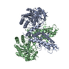
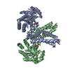
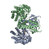
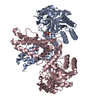
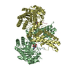
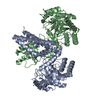
| Entry | Database: PDB / ID: 5m7t | |||||||||
|---|---|---|---|---|---|---|---|---|---|---|
| Title | Structure of human O-GlcNAc hydrolase with PugNAc type inhibitor | |||||||||
 Components Components | Protein O-GlcNAcase | |||||||||
 Keywords Keywords | HYDROLASE / human glacoside hydrolase / GlcNAc | |||||||||
| Function / homology |  Function and homology information Function and homology informationglycoprotein metabolic process / hyalurononglucosaminidase activity / N-acetylglucosamine metabolic process / glycoprotein catabolic process / protein O-GlcNAcase / : / : / [protein]-3-O-(N-acetyl-D-glucosaminyl)-L-serine/L-threonine O-N-acetyl-alpha-D-glucosaminase activity / protein O-linked glycosylation / protein deglycosylation ...glycoprotein metabolic process / hyalurononglucosaminidase activity / N-acetylglucosamine metabolic process / glycoprotein catabolic process / protein O-GlcNAcase / : / : / [protein]-3-O-(N-acetyl-D-glucosaminyl)-L-serine/L-threonine O-N-acetyl-alpha-D-glucosaminase activity / protein O-linked glycosylation / protein deglycosylation / beta-N-acetylglucosaminidase activity / membrane / identical protein binding / nucleus / cytosolSimilarity search - Function | |||||||||
| Biological species |  Homo sapiens (human) Homo sapiens (human) | |||||||||
| Method | X-RAY DIFFRACTION / SYNCHROTRON / MOLECULAR REPLACEMENT / Resolution: 2.6 Å | |||||||||
 Authors Authors | Roth, C. / Chan, S. / Offen, W.A. / Hemsworth, G.R. / Willems, L.I. / King, D. / Varghese, V. / Britton, R. / Vocadlo, D.J. / Davies, G.J. | |||||||||
| Funding support |  United Kingdom, United Kingdom,  Canada, 2items Canada, 2items
| |||||||||
 Citation Citation | Journal: Nat. Chem. Biol. / Year: 2017 Title: Structural and functional insight into human O-GlcNAcase. Authors: Roth, C. / Chan, S. / Offen, W.A. / Hemsworth, G.R. / Willems, L.I. / King, D.T. / Varghese, V. / Britton, R. / Vocadlo, D.J. / Davies, G.J. | |||||||||
| History |
|
- Structure visualization
| Structure viewer | Molecule: MolmilJmol/JSmol |
|---|
- Downloads & links
-Download
| PDBx/mmCIF format | 5m7t.cif.gz | 396.1 KB | Display | PDBx/mmCIF format |
|---|---|---|---|---|
| PDB format | pdb5m7t.ent.gz | 331.5 KB | Display | PDB format |
| PDBx/mmJSON format | 5m7t.json.gz | Tree view | PDBx/mmJSON format | |
| Others |  Other downloads Other downloads |
-Validation report
| Arichive directory | https://data.pdbj.org/pub/pdb/validation_reports/m7/5m7tftp://data.pdbj.org/pub/pdb/validation_reports/m7/5m7t | HTTPS FTP |
|---|
-Related structure data


-Links
 PDBj
PDBj


- Assembly
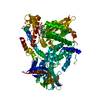
| Deposited unit | 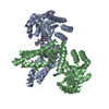
| ||||||||
|---|---|---|---|---|---|---|---|---|---|
| 1 |
| ||||||||
| Unit cell |
|
-Components
| #1: Protein | / OGA / Beta-N-acetylglucosaminidase / Beta-N-acetylhexosaminidase / Beta-hexosaminidase / Meningioma- ...OGA / Beta-N-acetylglucosaminidase / Beta-N-acetylhexosaminidase / Beta-hexosaminidase / Meningioma-expressed antigen 5 / N-acetyl-beta-D-glucosaminidase / N-acetyl-beta-glucosaminidase / Nuclear cytoplasmic O-GlcNAcase and acetyltransferase / NCOAT Mass: 103020.906 Da / Num. of mol.: 2 Source method: isolated from a genetically manipulated source Source: (gene. exp.) Homo sapiens (human) / Gene: MGEA5, HEXC, KIAA0679, MEA5 / Production host:  Escherichia coli BL21(DE3) (bacteria) / Variant (production host): Gold Escherichia coli BL21(DE3) (bacteria) / Variant (production host): GoldReferences: UniProt: O60502, protein O-GlcNAcase, Hydrolases; Glycosylases; Glycosidases, i.e. enzymes that hydrolyse O- and S-glycosyl compounds#2: Chemical |   Mass: 361.372 Da / Num. of mol.: 2 / Source method: obtained synthetically / Formula: C17H21N4O5 Mass: 361.372 Da / Num. of mol.: 2 / Source method: obtained synthetically / Formula: C17H21N4O5 |
|---|
-Experimental details
-Experiment
| Experiment | Method: X-RAY DIFFRACTION / Number of used crystals: 1 |
|---|
- Sample preparation
| Crystal | Density Matthews: 2.5 Å3/Da / Density % sol: 49.9 % |
|---|---|
| Crystal grow | Temperature: 292 K / Method: vapor diffusion / Details: 0.1-0.2 M tri Ammonium citrate 16-20 % PEG 3350 |
-Data collection
| Diffraction | Mean temperature: 100 K |
|---|---|
| Diffraction source | Source: SYNCHROTRON / Site: Diamond / Beamline: I02 / Wavelength: 0.979 Å |
| Detector | Type: DECTRIS PILATUS3 6M / Detector: PIXEL / Date: Aug 1, 2016 |
| Radiation | Protocol: SINGLE WAVELENGTH / Monochromatic (M) / Laue (L): M / Scattering type: x-ray |
| Radiation wavelength | Wavelength: 0.979 Å / Relative weight: 1 |
| Reflection | Resolution: 2.6→95.44 Å / Num. obs: 46333 / % possible obs: 100 % / Redundancy: 13.2 % / Rmerge(I) obs: 0.092 / Rpim(I) all: 0.026 / Net I/σ(I): 16.1 |
| Reflection shell | Resolution: 2.6→2.69 Å / Redundancy: 13.7 % / Rmerge(I) obs: 2.279 / Mean I/σ(I) obs: 1.1 / CC1/2: 0.59 / Rpim(I) all: 0.634 / % possible all: 100 |
- Processing
| Software |
| |||||||||||||||||||||||||||||||||||||||||||||||||||||||||||||||||||||||||||||||||||||||||||||||||||||||||||||||||||||||||||||||||||||||||||||||||||||||||||||||||||||||||||||||
|---|---|---|---|---|---|---|---|---|---|---|---|---|---|---|---|---|---|---|---|---|---|---|---|---|---|---|---|---|---|---|---|---|---|---|---|---|---|---|---|---|---|---|---|---|---|---|---|---|---|---|---|---|---|---|---|---|---|---|---|---|---|---|---|---|---|---|---|---|---|---|---|---|---|---|---|---|---|---|---|---|---|---|---|---|---|---|---|---|---|---|---|---|---|---|---|---|---|---|---|---|---|---|---|---|---|---|---|---|---|---|---|---|---|---|---|---|---|---|---|---|---|---|---|---|---|---|---|---|---|---|---|---|---|---|---|---|---|---|---|---|---|---|---|---|---|---|---|---|---|---|---|---|---|---|---|---|---|---|---|---|---|---|---|---|---|---|---|---|---|---|---|---|---|---|---|---|
| Refinement | Method to determine structure: MOLECULAR REPLACEMENT / Resolution: 2.6→95.44 Å / Cor.coef. Fo:Fc: 0.951 / Cor.coef. Fo:Fc free: 0.935 / Cross valid method: THROUGHOUT / σ(F): 0 / ESU R: 0.372 / ESU R Free: 0.251 Details: HYDROGENS HAVE BEEN ADDED IN THE RIDING POSITIONS U VALUES : WITH TLS ADDED
| |||||||||||||||||||||||||||||||||||||||||||||||||||||||||||||||||||||||||||||||||||||||||||||||||||||||||||||||||||||||||||||||||||||||||||||||||||||||||||||||||||||||||||||||
| Solvent computation | Ion probe radii: 0.8 Å / Shrinkage radii: 0.8 Å / VDW probe radii: 1.2 Å | |||||||||||||||||||||||||||||||||||||||||||||||||||||||||||||||||||||||||||||||||||||||||||||||||||||||||||||||||||||||||||||||||||||||||||||||||||||||||||||||||||||||||||||||
| Displacement parameters | Biso max: 218.95 Å2 / Biso mean: 105.8739 Å2 / Biso min: 50.68 Å2
| |||||||||||||||||||||||||||||||||||||||||||||||||||||||||||||||||||||||||||||||||||||||||||||||||||||||||||||||||||||||||||||||||||||||||||||||||||||||||||||||||||||||||||||||
| Refinement step | Cycle: LAST / Resolution: 2.6→95.44 Å
| |||||||||||||||||||||||||||||||||||||||||||||||||||||||||||||||||||||||||||||||||||||||||||||||||||||||||||||||||||||||||||||||||||||||||||||||||||||||||||||||||||||||||||||||
| Refine LS restraints |
| |||||||||||||||||||||||||||||||||||||||||||||||||||||||||||||||||||||||||||||||||||||||||||||||||||||||||||||||||||||||||||||||||||||||||||||||||||||||||||||||||||||||||||||||
| LS refinement shell | Resolution: 2.6→2.667 Å / Total num. of bins used: 20
| |||||||||||||||||||||||||||||||||||||||||||||||||||||||||||||||||||||||||||||||||||||||||||||||||||||||||||||||||||||||||||||||||||||||||||||||||||||||||||||||||||||||||||||||
| Refinement TLS params. | Method: refined / Refine-ID: X-RAY DIFFRACTION
| |||||||||||||||||||||||||||||||||||||||||||||||||||||||||||||||||||||||||||||||||||||||||||||||||||||||||||||||||||||||||||||||||||||||||||||||||||||||||||||||||||||||||||||||
| Refinement TLS group |
|
