| Entry | Database: PDB / ID: 5m6o
|
|---|
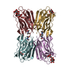
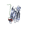
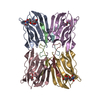
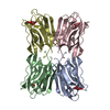
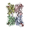
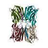
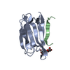
| Title | Frutapin complexed with alpha-D-mannose |
|---|
 Components Components | Frutapin |
|---|
 Keywords Keywords | carbohydrate-binding protein / Plant lectin /  Complex Complex |
|---|
| Function / homology |  Function and homology information Function and homology information |
|---|
| Biological species |  Artocarpus altilis (breadfruit) Artocarpus altilis (breadfruit) |
|---|
| Method | X-RAY DIFFRACTION / SYNCHROTRON / MOLECULAR REPLACEMENT / Resolution: 1.7 Å |
|---|
 Authors Authors | de Sousa, F.D. / Guo, J. / Coker, A.R. / de Oliveira Monteiro-Moreira, A. / de Azevedo Moreira, R. |
|---|
| Funding support |  Brazil, 1items Brazil, 1items | Organization | Grant number | Country |
|---|
| National Counsel of Technological and Scientific Development - CNPq | 201016/2015-0 | Brazil |
|
|---|
 Citation Citation | Journal: Biosci. Rep. / Year: 2017
Title: Frutapin, a lectin fromArtocarpus incisa(breadfruit): cloning, expression and molecular insights.
Authors: de Sousa, F.D. / da Silva, B.B. / Furtado, G.P. / Carneiro, I.S. / Lobo, M.D.P. / Guan, Y. / Guo, J. / Coker, A.R. / Lourenzoni, M.R. / Guedes, M.I.F. / Owen, J.S. / Abraham, D.J. / Monteiro- ...Authors: de Sousa, F.D. / da Silva, B.B. / Furtado, G.P. / Carneiro, I.S. / Lobo, M.D.P. / Guan, Y. / Guo, J. / Coker, A.R. / Lourenzoni, M.R. / Guedes, M.I.F. / Owen, J.S. / Abraham, D.J. / Monteiro-Moreira, A.C.O. / Moreira, R.A. |
|---|
| History | | Deposition | Oct 25, 2016 | Deposition site: PDBE / Processing site: PDBE |
|---|
| Revision 1.0 | Jul 19, 2017 | Provider: repository / Type: Initial release |
|---|
| Revision 1.1 | Aug 2, 2017 | Group: Database references / Category: citation / citation_author
Item: _citation.country / _citation.journal_volume / _citation_author.name |
|---|
| Revision 1.2 | Mar 28, 2018 | Group: Data collection / Database references / Category: citation / Item: _citation.title |
|---|
| Revision 1.3 | Jul 29, 2020 | Group: Data collection / Derived calculations / Structure summary
Category: chem_comp / entity ...chem_comp / entity / pdbx_chem_comp_identifier / pdbx_entity_nonpoly / struct_site / struct_site_gen
Item: _chem_comp.name / _chem_comp.type ..._chem_comp.name / _chem_comp.type / _entity.pdbx_description / _pdbx_entity_nonpoly.name
Description: Carbohydrate remediation / Provider: repository / Type: Remediation |
|---|
| Revision 1.4 | Jan 17, 2024 | Group: Data collection / Database references ...Data collection / Database references / Refinement description / Structure summary
Category: chem_comp / chem_comp_atom ...chem_comp / chem_comp_atom / chem_comp_bond / database_2 / pdbx_initial_refinement_model
Item: _chem_comp.pdbx_synonyms / _database_2.pdbx_DOI / _database_2.pdbx_database_accession |
|---|
|
|---|
 Movie
Movie Controller


 Open data
 Basic information
 Structure visualization
 Downloads & links
 Other downloads
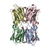


 PDBj
PDBj


 Assembly


 Type: D-saccharide, alpha linking / Mass: 180.156 Da / Num. of mol.: 4
 Mass: 18.015 Da / Num. of mol.: 427 / Source method: isolated from a natural source / Formula: H2O
 Sample preparation
 / Beamline: I24 / Wavelength: 0.96859 Å
 Processing