 Movie
Movie Controller

[English] 日本語
 Yorodumi
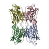
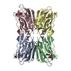
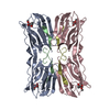
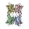
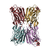
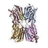
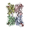
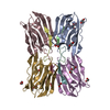
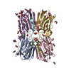
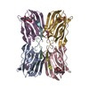
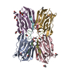
- PDB-1j4t: Structure of Artocarpin: a Lectin with Mannose Specificity (Form 2) -
+ Open data

- Basic information
| Entry | Database: PDB / ID: 1j4t | ||||||
|---|---|---|---|---|---|---|---|
| Title | Structure of Artocarpin: a Lectin with Mannose Specificity (Form 2) | ||||||
 Components Components | Artocarpin | ||||||
 Keywords Keywords |  PLANT PROTEIN / all Beta / Greek Key motif / Beta prism I fold PLANT PROTEIN / all Beta / Greek Key motif / Beta prism I fold | ||||||
| Function / homology |  Function and homology information Function and homology information | ||||||
| Biological species |   Artocarpus integer (campedak) Artocarpus integer (campedak) | ||||||
| Method | X-RAY DIFFRACTION / Resolution: 2.4 Å | ||||||
 Authors Authors | Pratap, J.V. / Jeyaprakash, A.A. / Rani, P.G. / Sekar, K. / Surolia, A. / Vijayan, M. | ||||||
 Citation Citation | Journal: J.Mol.Biol. / Year: 2002 Title: Crystal structures of artocarpin, a Moraceae lectin with mannose specificity, and its complex with methyl-alpha-D-mannose: implications to the generation of carbohydrate specificity. Authors: Pratap, J.V. / Jeyaprakash, A.A. / Rani, P.G. / Sekar, K. / Surolia, A. / Vijayan, M. | ||||||
| History |
|
- Structure visualization
| Structure viewer | Molecule: MolmilJmol/JSmol |
|---|
- Downloads & links
-Download
| PDBx/mmCIF format | 1j4t.cif.gz | 251.7 KB | Display | PDBx/mmCIF format |
|---|---|---|---|---|
| PDB format | pdb1j4t.ent.gz | 202.7 KB | Display | PDB format |
| PDBx/mmJSON format | 1j4t.json.gz | Tree view | PDBx/mmJSON format | |
| Others |  Other downloads Other downloads |
-Validation report
| Arichive directory | https://data.pdbj.org/pub/pdb/validation_reports/j4/1j4tftp://data.pdbj.org/pub/pdb/validation_reports/j4/1j4t | HTTPS FTP |
|---|
-Related structure data


-Links
 PDBj
PDBj- Assembly
| Deposited unit | 
| ||||||||
|---|---|---|---|---|---|---|---|---|---|
| 1 | 
| ||||||||
| 2 | 
| ||||||||
| Unit cell |
|
-Components
| #1: Protein | Mass: 16116.977 Da / Num. of mol.: 8 / Source method: isolated from a natural source / Details: seeds / Source: (natural) Artocarpus integer (campedak) / References: UniProt: Q7M1T4#2: Water | ChemComp-HOH / | Water Mass: 18.015 Da / Num. of mol.: 874 / Source method: isolated from a natural source / Formula: H2O Mass: 18.015 Da / Num. of mol.: 874 / Source method: isolated from a natural source / Formula: H2O |
|---|
-Experimental details
-Experiment
| Experiment | Method: X-RAY DIFFRACTION / Number of used crystals: 1 |
|---|
- Sample preparation
| Crystal | Density Matthews: 2.23 Å3/Da / Density % sol: 44.83 % | |||||||||||||||||||||||||||||||||||
|---|---|---|---|---|---|---|---|---|---|---|---|---|---|---|---|---|---|---|---|---|---|---|---|---|---|---|---|---|---|---|---|---|---|---|---|---|
| Crystal grow | Temperature: 293 K / Method: vapor diffusion / pH: 7.4 Details: PBS Buffer, 20% PEG 1450, 10Mg/ml protein, pH 7.4, vapor diffusion, temperature 293.0K | |||||||||||||||||||||||||||||||||||
| Crystal grow | *PLUS Method: vapor diffusion, hanging drop | |||||||||||||||||||||||||||||||||||
| Components of the solutions | *PLUS
|
-Data collection
| Diffraction | Mean temperature: 293 K |
|---|---|
| Diffraction source | Source: ROTATING ANODE / Wavelength: 1.5418 |
| Detector | Type: MARRESEARCH / Detector: IMAGE PLATE |
| Radiation | Protocol: SINGLE WAVELENGTH / Monochromatic (M) / Laue (L): M / Scattering type: x-ray |
| Radiation wavelength | Wavelength: 1.5418 Å / Relative weight: 1 |
| Reflection | Resolution: 2.4→20 Å / Num. all: 42342 / Num. obs: 42342 / % possible obs: 96.1 % / Redundancy: 3.1 % / Rmerge(I) obs: 0.089 |
| Reflection shell | Resolution: 2.4→2.5 Å / Redundancy: 3.2 % / Rmerge(I) obs: 0.379 / % possible all: 56 |
| Reflection | *PLUS Num. measured all: 133210 / Rmerge(I) obs: 0.089 |
| Reflection shell | *PLUS % possible obs: 56 % / Rmerge(I) obs: 0.379 |
- Processing
| Software |
| ||||||||||||||||||||||||
|---|---|---|---|---|---|---|---|---|---|---|---|---|---|---|---|---|---|---|---|---|---|---|---|---|---|
| Refinement | Resolution: 2.4→20 Å / σ(F): 0 / σ(I): 0 / Stereochemistry target values: Engh & Huber /
| ||||||||||||||||||||||||
| Refinement step | Cycle: LAST / Resolution: 2.4→20 Å
| ||||||||||||||||||||||||
| Refine LS restraints | Type: x_bond_d / Dev ideal: 0.01 | ||||||||||||||||||||||||
| Refinement | *PLUS Lowest resolution: 20 Å / Rfactor obs: 0.191 / Rfactor Rfree: 0.258 / Rfactor Rwork: 0.191 | ||||||||||||||||||||||||
| Solvent computation | *PLUS | ||||||||||||||||||||||||
| Displacement parameters | *PLUS | ||||||||||||||||||||||||
| Refine LS restraints | *PLUS
|
