 Movie
Movie Controller

[English] 日本語
 Yorodumi
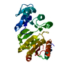
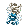
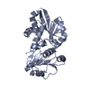
- PDB-5m08: Crystal structure of Mycobacterium tuberculosis PknI kinase domai... -
+ Open data

- Basic information
| Entry | Database: PDB / ID: 5m08 | ||||||
|---|---|---|---|---|---|---|---|
| Title | Crystal structure of Mycobacterium tuberculosis PknI kinase domain, C20A_R136A double mutant | ||||||
 Components Components | Serine/threonine-protein kinase PknI | ||||||
 Keywords Keywords |  SIGNALING PROTEIN / Tuberculosis / kinase / signalling SIGNALING PROTEIN / Tuberculosis / kinase / signalling | ||||||
| Function / homology |  Function and homology information Function and homology informationregulation of growth rate / peptidyl-threonine phosphorylation / manganese ion binding / peptidyl-serine phosphorylation / protein autophosphorylation / non-specific serine/threonine protein kinase / protein kinase activity / protein serine kinase activity / protein serine/threonine kinase activity / ATP binding ...regulation of growth rate / peptidyl-threonine phosphorylation / manganese ion binding / peptidyl-serine phosphorylation / protein autophosphorylation / non-specific serine/threonine protein kinase / protein kinase activity / protein serine kinase activity / protein serine/threonine kinase activity / ATP binding / plasma membrane / cytosolSimilarity search - Function | ||||||
| Biological species |   Mycobacterium tuberculosis (bacteria) Mycobacterium tuberculosis (bacteria) | ||||||
| Method | X-RAY DIFFRACTION / SYNCHROTRON / MOLECULAR REPLACEMENT / Resolution: 3.03 Å | ||||||
 Authors Authors | Lisa, M.N. / Wagner, T. / Alexandre, M. / Barilone, N. / Raynal, B. / Alzari, P.M. / Bellinzoni, M. | ||||||
 Citation Citation | Journal: FEBS J. / Year: 2017 Title: The crystal structure of PknI from Mycobacterium tuberculosis shows an inactive, pseudokinase-like conformation. Authors: Lisa, M.N. / Wagner, T. / Alexandre, M. / Barilone, N. / Raynal, B. / Alzari, P.M. / Bellinzoni, M. | ||||||
| History |
|
- Structure visualization
| Structure viewer | Molecule: MolmilJmol/JSmol |
|---|
- Downloads & links
-Download
| PDBx/mmCIF format | 5m08.cif.gz | 201.3 KB | Display | PDBx/mmCIF format |
|---|---|---|---|---|
| PDB format | pdb5m08.ent.gz | 162.1 KB | Display | PDB format |
| PDBx/mmJSON format | 5m08.json.gz | Tree view | PDBx/mmJSON format | |
| Others |  Other downloads Other downloads |
-Validation report
| Arichive directory | https://data.pdbj.org/pub/pdb/validation_reports/m0/5m08ftp://data.pdbj.org/pub/pdb/validation_reports/m0/5m08 | HTTPS FTP |
|---|
-Related structure data
| Related structure data |  5m06C  5m07SC  5m09C S: Starting model for refinement C: citing same article ( |
|---|---|
| Similar structure data |


-Links
 PDBj
PDBj- Assembly
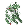
| Deposited unit | 
| ||||||||
|---|---|---|---|---|---|---|---|---|---|
| 1 | 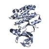
| ||||||||
| 2 | 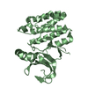
| ||||||||
| Unit cell |
|
-Components
| #1: Protein | Mass: 29552.297 Da / Num. of mol.: 2 / Mutation: C20A,R136A Source method: isolated from a genetically manipulated source Source: (gene. exp.) Mycobacterium tuberculosis (strain ATCC 25618 / H37Rv) (bacteria)Strain: ATCC 25618 / H37Rv / Gene: pknI, Rv2914c, MTCY338.02c / Production host: Escherichia coli BL21(DE3) (bacteria)References: UniProt: P9WI69, non-specific serine/threonine protein kinase#2: Water | ChemComp-HOH / | Water Mass: 18.015 Da / Num. of mol.: 22 / Source method: isolated from a natural source / Formula: H2O Mass: 18.015 Da / Num. of mol.: 22 / Source method: isolated from a natural source / Formula: H2O |
|---|
-Experimental details
-Experiment
| Experiment | Method: X-RAY DIFFRACTION / Number of used crystals: 1 |
|---|
- Sample preparation
| Crystal | Density Matthews: 4.65 Å3/Da / Density % sol: 73.56 % |
|---|---|
| Crystal grow | Temperature: 291 K / Method: vapor diffusion, hanging drop / pH: 7.5 / Details: NaCl 4.3 M, 0.1 M Hepes-Na pH 7.5 |
-Data collection
| Diffraction | Mean temperature: 100 K |
|---|---|
| Diffraction source | Source: SYNCHROTRON / Site: ESRF  / Beamline: ID29 / Wavelength: 0.97625 Å / Beamline: ID29 / Wavelength: 0.97625 Å |
| Detector | Type: DECTRIS PILATUS3 6M / Detector: PIXEL / Date: Oct 4, 2015 |
| Radiation | Protocol: SINGLE WAVELENGTH / Monochromatic (M) / Laue (L): M / Scattering type: x-ray |
| Radiation wavelength | Wavelength: 0.97625 Å / Relative weight: 1 |
| Reflection | Resolution: 3.03→48.88 Å / Num. obs: 22374 / % possible obs: 99.9 % / Redundancy: 5.4 % / Biso Wilson estimate: 96.2 Å2 / CC1/2: 0.999 / Rmerge(I) obs: 0.061 / Net I/σ(I): 9.7 |
| Reflection shell | Resolution: 3.03→3.21 Å / Redundancy: 5.4 % / Rmerge(I) obs: 0.444 / Mean I/σ(I) obs: 2 / CC1/2: 0.905 / % possible all: 100 |
- Processing
| Software |
| |||||||||||||||||||||||||||||||||||||||||||||||||||||||||||||||||||||||||||||||||||||||||||||||||||||||||||||||||||||||||||||
|---|---|---|---|---|---|---|---|---|---|---|---|---|---|---|---|---|---|---|---|---|---|---|---|---|---|---|---|---|---|---|---|---|---|---|---|---|---|---|---|---|---|---|---|---|---|---|---|---|---|---|---|---|---|---|---|---|---|---|---|---|---|---|---|---|---|---|---|---|---|---|---|---|---|---|---|---|---|---|---|---|---|---|---|---|---|---|---|---|---|---|---|---|---|---|---|---|---|---|---|---|---|---|---|---|---|---|---|---|---|---|---|---|---|---|---|---|---|---|---|---|---|---|---|---|---|---|
| Refinement | Method to determine structure: MOLECULAR REPLACEMENT Starting model: PDB entry 5M07 Resolution: 3.03→48.88 Å / Cor.coef. Fo:Fc: 0.9356 / Cor.coef. Fo:Fc free: 0.9189 / SU R Cruickshank DPI: 0.394 / Cross valid method: THROUGHOUT / σ(F): 0 / SU R Blow DPI: 0.394 / SU Rfree Blow DPI: 0.255 / SU Rfree Cruickshank DPI: 0.258
| |||||||||||||||||||||||||||||||||||||||||||||||||||||||||||||||||||||||||||||||||||||||||||||||||||||||||||||||||||||||||||||
| Displacement parameters | Biso mean: 87.18 Å2
| |||||||||||||||||||||||||||||||||||||||||||||||||||||||||||||||||||||||||||||||||||||||||||||||||||||||||||||||||||||||||||||
| Refine analyze | Luzzati coordinate error obs: 0.374 Å | |||||||||||||||||||||||||||||||||||||||||||||||||||||||||||||||||||||||||||||||||||||||||||||||||||||||||||||||||||||||||||||
| Refinement step | Cycle: 1 / Resolution: 3.03→48.88 Å
| |||||||||||||||||||||||||||||||||||||||||||||||||||||||||||||||||||||||||||||||||||||||||||||||||||||||||||||||||||||||||||||
| Refine LS restraints |
| |||||||||||||||||||||||||||||||||||||||||||||||||||||||||||||||||||||||||||||||||||||||||||||||||||||||||||||||||||||||||||||
| LS refinement shell | Resolution: 3.03→3.18 Å / Total num. of bins used: 11
| |||||||||||||||||||||||||||||||||||||||||||||||||||||||||||||||||||||||||||||||||||||||||||||||||||||||||||||||||||||||||||||
| Refinement TLS params. | Method: refined / Refine-ID: X-RAY DIFFRACTION
| |||||||||||||||||||||||||||||||||||||||||||||||||||||||||||||||||||||||||||||||||||||||||||||||||||||||||||||||||||||||||||||
| Refinement TLS group |
|
