 Movie
Movie Controller

[English] 日本語
 Yorodumi
- PDB-5lve: STRUCTURE OF THE VARIABLE DOMAIN OF HUMAN IMMUNOGLOBULIN K-4 LIGH... -
+ Open data

- Basic information
| Entry | Database: PDB / ID: 5lve | ||||||
|---|---|---|---|---|---|---|---|
| Title | STRUCTURE OF THE VARIABLE DOMAIN OF HUMAN IMMUNOGLOBULIN K-4 LIGHT CHAIN LEN | ||||||
 Components Components | BENCE-JONES PROTEIN LEN Bence Jones protein Bence Jones protein | ||||||
 Keywords Keywords | IMMUNE SYSTEM / IMMUNOGLOBULIN / BENCE-JONES PROTEIN | ||||||
| Function / homology |  Function and homology information Function and homology informationCD22 mediated BCR regulation / Fc epsilon receptor (FCERI) signaling / immunoglobulin complex / Classical antibody-mediated complement activation / Initial triggering of complement / FCGR activation / Role of phospholipids in phagocytosis / Role of LAT2/NTAL/LAB on calcium mobilization / Scavenging of heme from plasma / FCERI mediated Ca+2 mobilization ...CD22 mediated BCR regulation / Fc epsilon receptor (FCERI) signaling / immunoglobulin complex / Classical antibody-mediated complement activation / Initial triggering of complement / FCGR activation / Role of phospholipids in phagocytosis / Role of LAT2/NTAL/LAB on calcium mobilization / Scavenging of heme from plasma / FCERI mediated Ca+2 mobilization / FCGR3A-mediated IL10 synthesis / Antigen activates B Cell Receptor (BCR) leading to generation of second messengers / antigen binding / Regulation of Complement cascade / Cell surface interactions at the vascular wall / FCERI mediated MAPK activation / FCGR3A-mediated phagocytosis / Regulation of actin dynamics for phagocytic cup formation / FCERI mediated NF-kB activation / Immunoregulatory interactions between a Lymphoid and a non-Lymphoid cell / adaptive immune response / Potential therapeutics for SARS / blood microparticle / immune response / extracellular space / extracellular region / plasma membraneSimilarity search - Function | ||||||
| Biological species |  Homo sapiens (human) Homo sapiens (human) | ||||||
| Method | X-RAY DIFFRACTION / MOLECULAR REPLACEMENT / Resolution: 2 Å | ||||||
 Authors Authors | Schiffer, M. / Huang, D.-B. / Chang, C.-H. | ||||||
 Citation Citation | Journal: Protein Sci. / Year: 2000 Title: Change in dimerization mode by removal of a single unsatisfied polar residue located at the interface. Authors: Pokkuluri, P.R. / Cai, X. / Johnson, G. / Stevens, F.J. / Schiffer, M. #1: Journal: Structure / Year: 1998Title: A domain flip as a result of a single amino-acid substitution. Authors: Pokkuluri, P.R. / Huang, D.B. / Raffen, R. / Cai, X. / Johnson, G. / Stevens, P.W. / Stevens, F.J. / Schiffer, M. | ||||||
| History |
|
- Structure visualization
| Structure viewer | Molecule: MolmilJmol/JSmol |
|---|
- Downloads & links
-Download
| PDBx/mmCIF format | 5lve.cif.gz | 40.1 KB | Display | PDBx/mmCIF format |
|---|---|---|---|---|
| PDB format | pdb5lve.ent.gz | 26.5 KB | Display | PDB format |
| PDBx/mmJSON format | 5lve.json.gz | Tree view | PDBx/mmJSON format | |
| Others |  Other downloads Other downloads |
-Validation report
| Arichive directory | https://data.pdbj.org/pub/pdb/validation_reports/lv/5lveftp://data.pdbj.org/pub/pdb/validation_reports/lv/5lve | HTTPS FTP |
|---|
-Related structure data
| Related structure data | 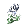 1qacC 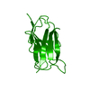 1lveS S: Starting model for refinement C: citing same article ( |
|---|---|
| Similar structure data |


-Links
 PDBj
PDBj


- Assembly
| Deposited unit | 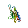
| |||||||||||||||
|---|---|---|---|---|---|---|---|---|---|---|---|---|---|---|---|---|
| 1 | 
| |||||||||||||||
| 2 | 
| |||||||||||||||
| Unit cell |
| |||||||||||||||
| Components on special symmetry positions |
|
-Components
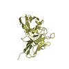
| #1: Antibody | Bence Jones protein Mass: 12592.961 Da / Num. of mol.: 1 / Fragment: G KAPPA CHAIN V-IV, VARIABLE DOMAIN, LIGHT CHAIN / Mutation: Q89A Source method: isolated from a genetically manipulated source Source: (gene. exp.) Homo sapiens (human) / Production host:  Escherichia coli (E. coli) / Strain (production host): JM83 / References: UniProt: P01625, UniProt: P06312*PLUS Escherichia coli (E. coli) / Strain (production host): JM83 / References: UniProt: P01625, UniProt: P06312*PLUS |
|---|---|
| #2: Chemical | ChemComp-ZN /   Mass: 65.409 Da / Num. of mol.: 1 / Source method: obtained synthetically / Formula: Zn Mass: 65.409 Da / Num. of mol.: 1 / Source method: obtained synthetically / Formula: Zn |
| #3: Water | ChemComp-HOH / Water Mass: 18.015 Da / Num. of mol.: 156 / Source method: isolated from a natural source / Formula: H2O Mass: 18.015 Da / Num. of mol.: 156 / Source method: isolated from a natural source / Formula: H2O |
| Nonpolymer details | A ZN+2 ION (RESIDUE 200) AND THREE WATERS (RESIDUES 214, 274 AND 353) ARE LOCATED ON SYMMETRY AXES ...A ZN+2 ION (RESIDUE 200) AND THREE WATERS (RESIDUES 214, 274 AND 353) ARE LOCATED ON SYMMETRY AXES AND THEIR OCCUPANCY IS SET TO 0.5 |
| Sequence details | Q89A MUTANT |
-Experimental details
-Experiment
| Experiment | Method: X-RAY DIFFRACTION / Number of used crystals: 1 |
|---|
- Sample preparation
| Crystal | Density Matthews: 3 Å3/Da / Density % sol: 59 % | |||||||||||||||||||||||||
|---|---|---|---|---|---|---|---|---|---|---|---|---|---|---|---|---|---|---|---|---|---|---|---|---|---|---|
| Crystal grow | pH: 7.5 Details: 2.0 M AMMONIUM SULFATE, 0.1M HEPES PH 7.5, 1MM ZINC ACETATE | |||||||||||||||||||||||||
| Crystal grow | *PLUS Method: vapor diffusion, hanging drop | |||||||||||||||||||||||||
| Components of the solutions | *PLUS
|
-Data collection
| Diffraction | Mean temperature: 100 K |
|---|---|
| Diffraction source | Source: ROTATING ANODE / Type: RIGAKU RU200 / Wavelength: 1.54 |
| Detector | Type: RIGAKU / Detector: IMAGE PLATE / Date: Jun 1, 1998 / Details: MIRRORS |
| Radiation | Monochromator: NI FILTER / Protocol: SINGLE WAVELENGTH / Monochromatic (M) / Laue (L): M / Scattering type: x-ray |
| Radiation wavelength | Wavelength: 1.54 Å / Relative weight: 1 |
| Reflection | Resolution: 2→30 Å / Num. obs: 7471 / % possible obs: 97 % / Redundancy: 10 % / Biso Wilson estimate: 6.6 Å2 / Rmerge(I) obs: 0.074 / Net I/σ(I): 25 |
| Reflection shell | Resolution: 2→2.07 Å / Redundancy: 15.7 % / Rmerge(I) obs: 0.242 / Mean I/σ(I) obs: 6.8 / % possible all: 88.3 |
| Reflection shell | *PLUS % possible obs: 88.3 % |
- Processing
| Software |
| ||||||||||||||||||||||||||||||||||||||||||||||||||||||||||||||||||||||||||||||||
|---|---|---|---|---|---|---|---|---|---|---|---|---|---|---|---|---|---|---|---|---|---|---|---|---|---|---|---|---|---|---|---|---|---|---|---|---|---|---|---|---|---|---|---|---|---|---|---|---|---|---|---|---|---|---|---|---|---|---|---|---|---|---|---|---|---|---|---|---|---|---|---|---|---|---|---|---|---|---|---|---|---|
| Refinement | Method to determine structure: MOLECULAR REPLACEMENT Starting model: 1LVE WITH GLN89 CHANGED TO ALA Resolution: 2→8 Å / Rfactor Rfree error: 0.007 / Data cutoff high rms absF: 388280.78 / Isotropic thermal model: RESTRAINED / Cross valid method: THROUGHOUT / σ(F): 3 Details: BULK SOLVENT MODEL USED THE LAST TWO RESIDUES (LYS-107 AND ARG-108) ARE NOT WELL DEFINED IN THE DENSITY MAPS.
| ||||||||||||||||||||||||||||||||||||||||||||||||||||||||||||||||||||||||||||||||
| Solvent computation | Solvent model: FLAT MODEL / Bsol: 73.8 Å2 / ksol: 0.5 e/Å3 | ||||||||||||||||||||||||||||||||||||||||||||||||||||||||||||||||||||||||||||||||
| Displacement parameters | Biso mean: 18.6 Å2
| ||||||||||||||||||||||||||||||||||||||||||||||||||||||||||||||||||||||||||||||||
| Refine analyze |
| ||||||||||||||||||||||||||||||||||||||||||||||||||||||||||||||||||||||||||||||||
| Refinement step | Cycle: LAST / Resolution: 2→8 Å
| ||||||||||||||||||||||||||||||||||||||||||||||||||||||||||||||||||||||||||||||||
| Refine LS restraints |
| ||||||||||||||||||||||||||||||||||||||||||||||||||||||||||||||||||||||||||||||||
| LS refinement shell | Resolution: 2→2.04 Å / Rfactor Rfree error: 0.031 / Total num. of bins used: 18
| ||||||||||||||||||||||||||||||||||||||||||||||||||||||||||||||||||||||||||||||||
| Xplor file |
| ||||||||||||||||||||||||||||||||||||||||||||||||||||||||||||||||||||||||||||||||
| Software | *PLUS Name: CNS / Version: 0.3 / Classification: refinement | ||||||||||||||||||||||||||||||||||||||||||||||||||||||||||||||||||||||||||||||||
| Refine LS restraints | *PLUS
|
