 Movie
Movie Controller

[English] 日本語
 Yorodumi
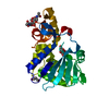
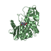
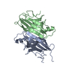
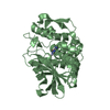
- PDB-1qac: CHANGE IN DIMERIZATION MODE BY REMOVAL OF A SINGLE UNSATISFIED PO... -
+ Open data

- Basic information
| Entry | Database: PDB / ID: 1qac | ||||||
|---|---|---|---|---|---|---|---|
| Title | CHANGE IN DIMERIZATION MODE BY REMOVAL OF A SINGLE UNSATISFIED POLAR RESIDUE | ||||||
 Components Components | IMMUNOGLOBULIN LIGHT CHAIN VARIABLE DOMAIN | ||||||
 Keywords Keywords |  IMMUNE SYSTEM / BETA BARREL IMMUNOGLOBULIN VL DOMAIN DIMER / FLIPPED DOMAIN DIMER IMMUNE SYSTEM / BETA BARREL IMMUNOGLOBULIN VL DOMAIN DIMER / FLIPPED DOMAIN DIMER | ||||||
| Function / homology |  Function and homology information Function and homology informationCD22 mediated BCR regulation / Fc epsilon receptor (FCERI) signaling / immunoglobulin complex / Classical antibody-mediated complement activation / Initial triggering of complement / FCGR activation / Role of phospholipids in phagocytosis / Role of LAT2/NTAL/LAB on calcium mobilization / Scavenging of heme from plasma / FCERI mediated Ca+2 mobilization ...CD22 mediated BCR regulation / Fc epsilon receptor (FCERI) signaling / immunoglobulin complex / Classical antibody-mediated complement activation / Initial triggering of complement / FCGR activation / Role of phospholipids in phagocytosis / Role of LAT2/NTAL/LAB on calcium mobilization / Scavenging of heme from plasma / FCERI mediated Ca+2 mobilization / FCGR3A-mediated IL10 synthesis / Antigen activates B Cell Receptor (BCR) leading to generation of second messengers / antigen binding / Regulation of Complement cascade / Cell surface interactions at the vascular wall / FCERI mediated MAPK activation / FCGR3A-mediated phagocytosis / Regulation of actin dynamics for phagocytic cup formation / FCERI mediated NF-kB activation / Immunoregulatory interactions between a Lymphoid and a non-Lymphoid cell / adaptive immune response / Potential therapeutics for SARS / blood microparticle / immune response / extracellular space / extracellular region / plasma membraneSimilarity search - Function | ||||||
| Biological species |  Homo sapiens (human) Homo sapiens (human) | ||||||
| Method | X-RAY DIFFRACTION / SYNCHROTRON / MOLECULAR REPLACEMENT / Resolution: 1.8 Å | ||||||
 Authors Authors | Pokkuluri, P.R. / Cai, X. / Johnson, G. / Stevens, F.J. / Schiffer, M. | ||||||
 Citation Citation | Journal: Protein Sci. / Year: 2000 Title: Change in dimerization mode by removal of a single unsatisfied polar residue located at the interface. Authors: Pokkuluri, P.R. / Cai, X. / Johnson, G. / Stevens, F.J. / Schiffer, M. #1: Journal: Structure / Year: 1998Title: A domain flip as a result of a single amino-acid substitution. Authors: Pokkuluri, P.R. / Huang, D.-B. / Raffen, R. / Cai, X. / Johnson, G. / Wilkins Stevens, P. / Stevens, F.J. / Schiffer, M. | ||||||
| History |
|
- Structure visualization
| Structure viewer | Molecule: MolmilJmol/JSmol |
|---|
- Downloads & links
-Download
| PDBx/mmCIF format | 1qac.cif.gz | 63.9 KB | Display | PDBx/mmCIF format |
|---|---|---|---|---|
| PDB format | pdb1qac.ent.gz | 45.3 KB | Display | PDB format |
| PDBx/mmJSON format | 1qac.json.gz | Tree view | PDBx/mmJSON format | |
| Others |  Other downloads Other downloads |
-Validation report
| Arichive directory | https://data.pdbj.org/pub/pdb/validation_reports/qa/1qacftp://data.pdbj.org/pub/pdb/validation_reports/qa/1qac | HTTPS FTP |
|---|
-Related structure data
| Related structure data |  5lveC 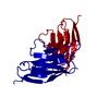 3lveS S: Starting model for refinement C: citing same article ( |
|---|---|
| Similar structure data |


-Links
 PDBj
PDBj


- Assembly
| Deposited unit | 
| ||||||||||
|---|---|---|---|---|---|---|---|---|---|---|---|
| 1 |
| ||||||||||
| Unit cell |
|
-Components
| #1: Antibody | Mass: 12635.041 Da / Num. of mol.: 2 / Fragment: VARIABLE DOMAIN / Mutation: Q89L Source method: isolated from a genetically manipulated source Source: (gene. exp.) Homo sapiens (human) / Production host:  Escherichia coli (E. coli) / References: UniProt: P06312*PLUS Escherichia coli (E. coli) / References: UniProt: P06312*PLUS#2: Water | ChemComp-HOH / | Water Mass: 18.015 Da / Num. of mol.: 305 / Source method: isolated from a natural source / Formula: H2O Mass: 18.015 Da / Num. of mol.: 305 / Source method: isolated from a natural source / Formula: H2O |
|---|
-Experimental details
-Experiment
| Experiment | Method: X-RAY DIFFRACTION / Number of used crystals: 1 |
|---|
- Sample preparation
| Crystal | Density Matthews: 2.6 Å3/Da / Density % sol: 53 % | |||||||||||||||
|---|---|---|---|---|---|---|---|---|---|---|---|---|---|---|---|---|
| Crystal grow | Temperature: 277 K / Method: vapor diffusion Details: 18% PEG 8000, 0.2M AMMONIUM SULPHATE AT 4 DEG C, VAPOR DIFFUSION, temperature 277K | |||||||||||||||
| Crystal grow | *PLUS Method: vapor diffusion, hanging drop | |||||||||||||||
| Components of the solutions | *PLUS
|
-Data collection
| Diffraction | Mean temperature: 100 K |
|---|---|
| Diffraction source | Source: SYNCHROTRON / Site: APS  / Beamline: 19-ID / Wavelength: 1.033 / Beamline: 19-ID / Wavelength: 1.033 |
| Detector | Type: CUSTOM-MADE / Detector: CCD / Date: Nov 29, 1998 |
| Radiation | Protocol: SINGLE WAVELENGTH / Monochromatic (M) / Laue (L): M / Scattering type: x-ray |
| Radiation wavelength | Wavelength: 1.033 Å / Relative weight: 1 |
| Reflection | Resolution: 1.8→20 Å / Num. all: 22265 / Num. obs: 21776 / % possible obs: 97.9 % / Observed criterion σ(I): 1 / Redundancy: 8.6 % / Biso Wilson estimate: 8 Å2 / Rmerge(I) obs: 0.056 / Net I/σ(I): 21.9 |
| Reflection shell | Resolution: 1.8→1.86 Å / Redundancy: 9.2 % / Rmerge(I) obs: 0.172 / Mean I/σ(I) obs: 5.6 |
- Processing
| Software |
| ||||||||||||||||||||||||||||||||||||||||||||||||||||||||||||||||||||||||||||||||
|---|---|---|---|---|---|---|---|---|---|---|---|---|---|---|---|---|---|---|---|---|---|---|---|---|---|---|---|---|---|---|---|---|---|---|---|---|---|---|---|---|---|---|---|---|---|---|---|---|---|---|---|---|---|---|---|---|---|---|---|---|---|---|---|---|---|---|---|---|---|---|---|---|---|---|---|---|---|---|---|---|---|
| Refinement | Method to determine structure: MOLECULAR REPLACEMENT Starting model: 3LVE with GLU-38 and GLN-89 changed to ALA Resolution: 1.8→8 Å / Rfactor Rfree error: 0.005 / Data cutoff high rms absF: 10000 / Data cutoff low absF: 0 / Isotropic thermal model: RESTRAINED / Cross valid method: THROUGHOUT / σ(F): 3 / Stereochemistry target values: ENGH & HUBER Details: CONJUGATE-GRADIENT MINIMIZATION. MAXIMUM LIKELIHOOD BASED ON AMPLITUDES TARGET USED
| ||||||||||||||||||||||||||||||||||||||||||||||||||||||||||||||||||||||||||||||||
| Solvent computation | Solvent model: FLAT / Bsol: 65.1 Å2 / ksol: 0.46 e/Å3 | ||||||||||||||||||||||||||||||||||||||||||||||||||||||||||||||||||||||||||||||||
| Displacement parameters | Biso mean: 16.9 Å2
| ||||||||||||||||||||||||||||||||||||||||||||||||||||||||||||||||||||||||||||||||
| Refine analyze |
| ||||||||||||||||||||||||||||||||||||||||||||||||||||||||||||||||||||||||||||||||
| Refinement step | Cycle: LAST / Resolution: 1.8→8 Å
| ||||||||||||||||||||||||||||||||||||||||||||||||||||||||||||||||||||||||||||||||
| Refine LS restraints |
| ||||||||||||||||||||||||||||||||||||||||||||||||||||||||||||||||||||||||||||||||
| LS refinement shell | Resolution: 1.8→1.83 Å / Rfactor Rfree error: 0.028 / Total num. of bins used: 18
| ||||||||||||||||||||||||||||||||||||||||||||||||||||||||||||||||||||||||||||||||
| Xplor file |
| ||||||||||||||||||||||||||||||||||||||||||||||||||||||||||||||||||||||||||||||||
| Software | *PLUS Name: CNS / Version: 0.3 / Classification: refinement | ||||||||||||||||||||||||||||||||||||||||||||||||||||||||||||||||||||||||||||||||
| Refinement | *PLUS Num. reflection all: 19237 | ||||||||||||||||||||||||||||||||||||||||||||||||||||||||||||||||||||||||||||||||
| Solvent computation | *PLUS | ||||||||||||||||||||||||||||||||||||||||||||||||||||||||||||||||||||||||||||||||
| Displacement parameters | *PLUS | ||||||||||||||||||||||||||||||||||||||||||||||||||||||||||||||||||||||||||||||||
| Refine LS restraints | *PLUS
|
