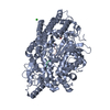
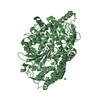
Entry Database : PDB / ID : 5lv0Title Structure of Human Neurolysin (E475Q) in complex with amyloid-beta 35-40 peptide product GLY-VAL-VAL amyloid 35-40 fragment Neurolysin, mitochondrial Keywords / / / Function / homology Function Domain/homology Component
/ / / / / / / / / / / / / / / / / / / / / / / / / / / / / / / / / / / / / / / / / / / / / / / / / / / / / / / / / / / / / / / / / / / / / / / / / / / / / / / / / / / / / / / / / / / / / / / / / / / / / / / / / / / / / / / / / / / / / / / / / / / / / / / / / / / / / / / / / / / / / / / / / / / / / / / / / / / / / / / / / / / / / Biological species Homo sapiens (human)Method / / / Resolution : 2.7 Å Authors Masuyer, G. / Berntsson, R.P.-A. / Teixeira, P.F. / Kmiec, B. / Glaser, E. / Stenmark, P. Funding support Organization Grant number Country Swedish Research Council 2014-5667
Journal : To Be Published Title : Structural and functional analysis of Neurolysin, a new component of the mitochondrial peptidolytic networkAuthors : Teixeira, P.F. / Masuyer, G. / Pinho, C. / Branca, R.M.M. / Kmiec, B. / Wallin, C. / Warmlander, S. / Berntsson, R.P.-A. / Ankarcrona, M. / Graslund, A. / Lehtio, J. / Stenmark, P. / Glaser, E. History Deposition Sep 12, 2016 Deposition site / Processing site Revision 1.0 Dec 6, 2017 Provider / Type Revision 1.1 Feb 7, 2018 Group / Structure summary / Category / citation_author / Item / _citation_author.nameRevision 1.2 Jan 17, 2024 Group Data collection / Database references ... Data collection / Database references / Derived calculations / Refinement description Category chem_comp_atom / chem_comp_bond ... chem_comp_atom / chem_comp_bond / database_2 / pdbx_initial_refinement_model / struct_conn Item _database_2.pdbx_DOI / _database_2.pdbx_database_accession ... _database_2.pdbx_DOI / _database_2.pdbx_database_accession / _struct_conn.pdbx_dist_value / _struct_conn.ptnr1_auth_asym_id / _struct_conn.ptnr1_auth_comp_id / _struct_conn.ptnr1_auth_seq_id / _struct_conn.ptnr1_label_asym_id / _struct_conn.ptnr1_label_atom_id / _struct_conn.ptnr1_label_comp_id / _struct_conn.ptnr1_label_seq_id / _struct_conn.ptnr2_auth_asym_id / _struct_conn.ptnr2_auth_comp_id / _struct_conn.ptnr2_auth_seq_id / _struct_conn.ptnr2_label_asym_id / _struct_conn.ptnr2_label_atom_id / _struct_conn.ptnr2_label_comp_id / _struct_conn.ptnr2_label_seq_id
Show all Show less
 Movie
Movie Controller

 Yorodumi
 Open data
 Basic information
 Components
 Keywords
 HYDROLASE /
  Function and homology information
 Authors
 Sweden, 1items
  Citation
 Structure visualization
 Downloads & links
 Other downloads


 PDBj
PDBj


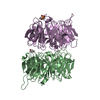
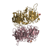
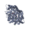
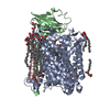
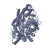
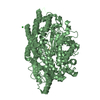
 Assembly


 Mass: 65.409 Da / Num. of mol.: 2 / Source method: obtained synthetically / Formula: Zn
 Mass: 35.453 Da / Num. of mol.: 2 / Source method: obtained synthetically / Formula: Cl
 Mass: 18.015 Da / Num. of mol.: 59 / Source method: isolated from a natural source / Formula: H2O
 Sample preparation
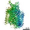
 / Beamline: P13 (MX1) / Wavelength: 0.976 Å
 Processing